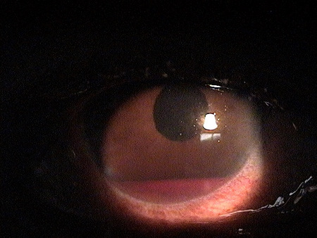
H21.01-03 Hyphema of Iris and Ciliary Body
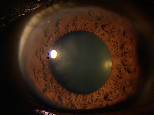
Hyphema is a condition where blood from the iris or ciliary body hemorrhages into the anterior chamber. A hyphema may appear as a few red blood cells or it can completely fill the anterior chamber.


Hyphema is a condition where blood from the iris or ciliary body hemorrhages into the anterior chamber. A hyphema may appear as a few red blood cells or it can completely fill the anterior chamber.
Iridocyclitis is an inflammation of the uvela tissues, primarily the iris and ciliary body.
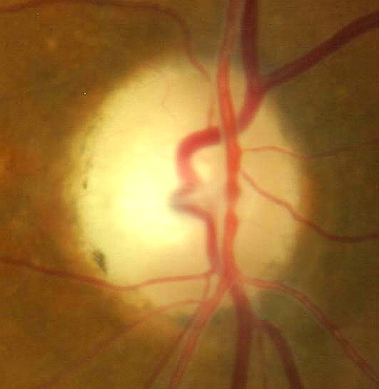
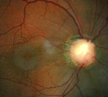
Partial optic atrophy is not a diagnosis in itself, but rather a clinical sign of a previous optic nerve condition.
A corneal erosion is characterized by a spontaneous detachment of the basal cells of the corneal epithelium from the underlying basement membrane.
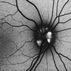
A condition involving superficial or buried calcium-like globular deposits anterior to the lamina cribrosa of the optic nerve head.
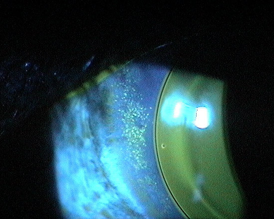
Contact lens wear can cause a change in corneal physiology, which may lead to lead to epithelial, stromal, and endothelial compromise.
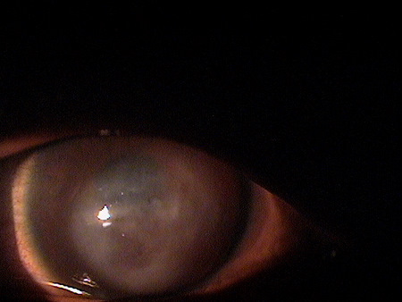
Descemetocele involves the anterior bulging or rupture of an intact Descemet's membrane through a defect of the overlying corneal stroma and epithelium.
Optic pits are excavations of the optic nerve head.
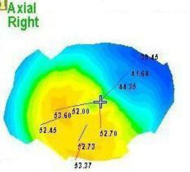
Pellucid marginal degeneration is a condition characterized by peripheral corneal thinning and corneal ectasia.
Corneal hydrops occurs after a rupture in Descemet's membrane and is characterized by severe corneal edema.