

 Print | Share
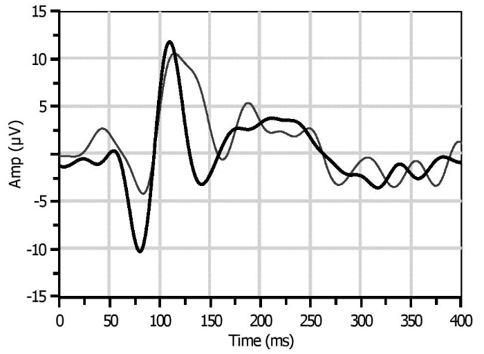
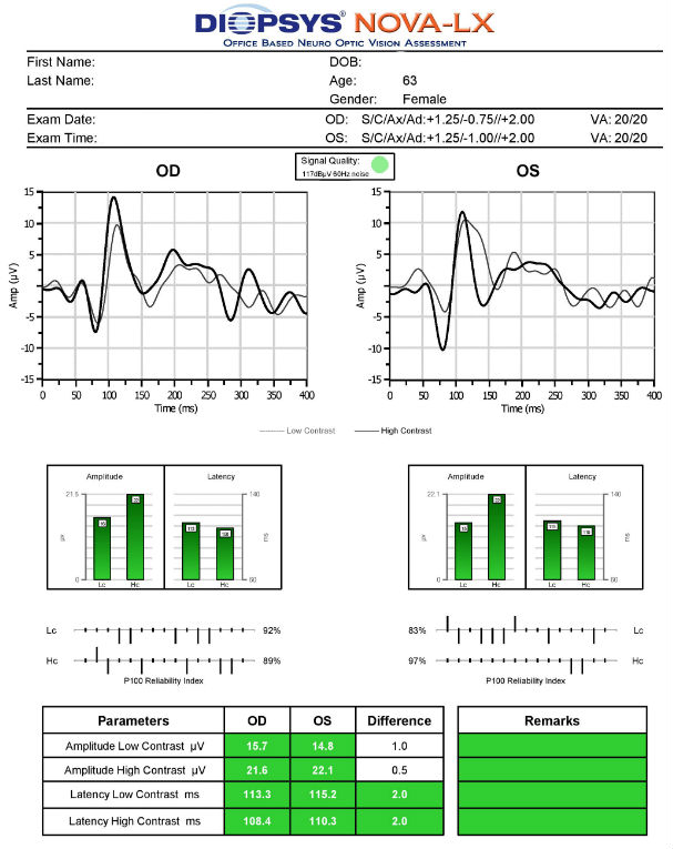
Normal VEP waveform in a glaucoma suspect
ICD-10 Diagnosis Codes:
H40.011–Open-angle glaucoma with borderline findings, low risk, right eye
H40.012–Open-angle glaucoma with borderline findings, low risk, left eye
H40.013–Open-angle glaucoma with borderline findings, low risk, bilateral
H40.021–Open-angle glaucoma with borderline findings, high risk, right eye
H40.022–Open-Angle Glaucoma with borderline findings, high risk, left eye
H40.023–Open-Angle Glaucoma with borderline findings, high risk, bilateral
Title
Open-Angle Glaucoma Suspect, low risk
Category
Glaucoma
Description
A glaucoma suspect is a person with clinical findings and/or a combination of risk factors that indicate an increased likelihood of developing glaucoma.
Glaucoma is an optic neuropathy showing distinctive changes in optic nerve morphology without associated pallor. The term “glaucoma” refers to a group of chronic, progressive optic neuropathies that have in common characteristic morphologic changes at the optic nerve and retinal nerve fiber layer.
The glaucomas are associated with the following clinical features:
- Aqueous outflow restrictions
- Unphysiologic intraocular pressure
- Abnormal ocular perfusion
- Abnormal rate of apoptosis
- Progressive retinal ganglion cell loss
- Characteristic changes in optic nerve anatomy
Conclusion
Glaucoma is a diagnosis of exclusion. According to the 2015 ICD-10 diagnosis codes, persons are considered open-angle glaucoma suspects based on the number of risk factors they possess. Low-risk is one or two risk factors. High-risk is three or more risk factors. Commonly considered risk factors include:
- Positive family history for glaucoma
- Black or Hispanic race
- Elevated intraocular pressure
- Abnormal optic disc appearance (esp. vertical C/D changes)
- Thin central corneal thickness below 500 microns combined with elevated intraocular pressures
Additional risk factors not included in the ICD-10 listing include the following:
- Low blood pressure
- Sleeping / breathing disorders
- Diabetes
- Angle abnormalities / outflow restrictions
- Prior trauma to globe
For patients diagnosed as glaucoma suspects, there remains some controversy on whether unphysiologic intraocular pressure is the dominant factor involved in developing glaucomatous optic atrophy or if there is a secondary component such as compromised blood flow to the optic nerve head.
 |
|
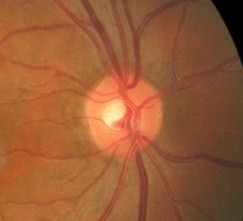
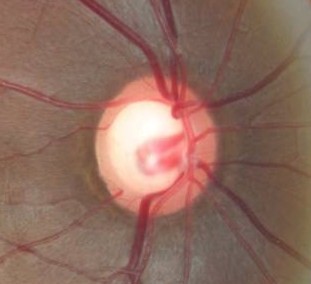
Normal Neuroretinal Rim Tissue
|
 |
 |
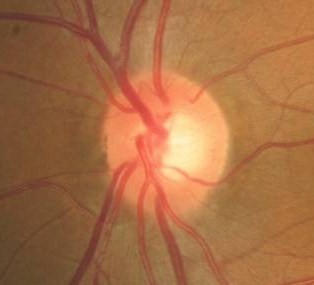
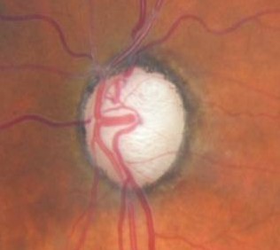
Changes in Coloration of the Optic Nerve
|
 |
Changes in Coloration of the Optic Nerve
|
 |
|
 |
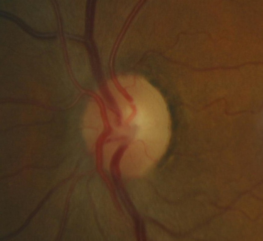
Changes in the Margins of the Optic Cup
|
 |
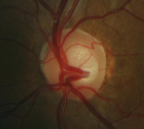
Changes in Optic Cup Depth
|
 |
|
 |
|
 |
Vessel Changes at the Optic Nerve
|
 |
Loss of Retinal Ganglion Cells
|
Functional Damage to the Visual System
1. Associated visual field defects
- Reduction in retinal sensitivity
- Constriction of isopters
- Nasal steps
- Paracentral scotomas
- Arcuate scotomas
- Hemifield asymmetry
- Temporal changes in visual field patterns
- Loss of central acuity
2. Decreased color vision
3. Abnormal visual evoked potential
4. Abnormal pattern electroretinogram
The main goal in a patient diagnosed as a glaucoma suspect is to accomplish the following:
- Identify patients at high-risk for developing glaucoma
- Establish a baseline of clinical findings for future comparative analysis
- Identify and exclude any oculosystemic conditions associated with open-angle glaucoma
- Consider medical treatment of high-risk patients to prevent or slow down the development of open-angle glaucoma
To obtain the information required to determine a clinical diagnosis of an open angle glaucoma suspect and prescribe a treatment program, the following service components of a medical eye examination should be performed:
- Patient history
- General medical observation
- Gross visual field
- Pupillary examination
- External ocular examination with biomicroscopy
- Ophthalmoscopy
Patient History
- Patients will usually be asymptomatic in the early stages of glaucoma
Intraocular Pressure
- Measurements above 21 mm Hg
- Asymmetry above 5 mm Hg
Gross Visual Fields
- Normal
- Constrictions
Pupillary Examination
- A relative afferent pupillary defect (RAPD) may be present if one eye is more affected than the other
External Ocular Examination with Biomicroscopy
- Normal
Ophthalmoscopic Examination
- Cup-to-disc ratio > .40/.40
- Cup-to-disc ratio asymmetry > .20/.20
- Vertically elongated cup-to-disc ratios
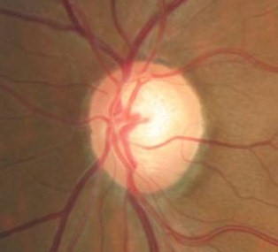
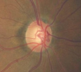
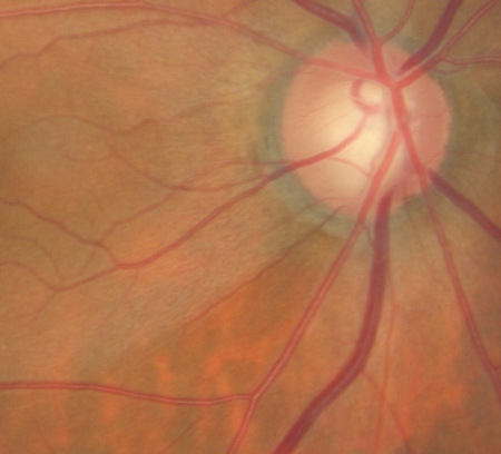
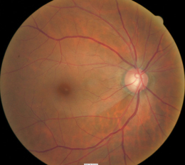
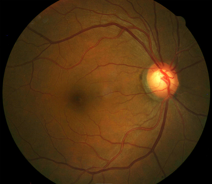
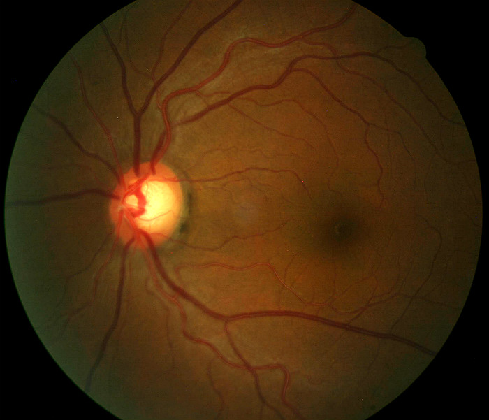
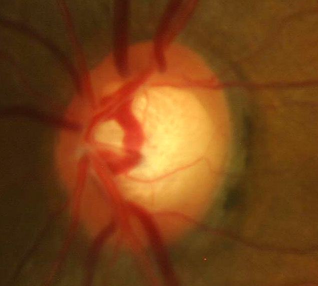
Patient is a 64-year-old black female with an optic disc appearance that is suspicious for glaucomatous optic atrophy.
- Cup-to-disc ratio = .75/.70 in the right eye
- Cup-to-disc ratio = .75/.70 in the left eye
- Normal neuroretinal rim
- Normal retinal nerve fiber layer
 |
 |
DIAGNOSTIC TESTS
The following diagnostic tests can provide clincial information in the evaluation of glaucoma and its stage of damage.
Retinal Scanning Laser
- Can show attenuation of the retinal nerve fiber layer
- Excavation of the optic disc
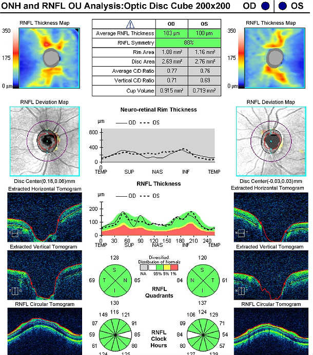 |
Cirrus OCT Scan in a 64-year-old Glaucoma Suspect
Left Eye
Both Eyes
|
Visual Field Examination
Research has shown that over 50% of the nerve fibers in any given retinal nerve fiber layer bundle can be irreversibly damaged before any visual field loss occurs. Depending upon visual fields to separate those patients with glaucoma from those without the disease would still miss a large number of patients.
For most patients with glaucoma, there appears to be about a five year interval between the loss of enough nerve fibers to create a visual field defect during a visual field examination. Relying on visual fields alone to diagnose glaucoma could mean delayed treatment for your glaucoma patients and unnecessary loss of visual field in these people.
Visual field defects will depend on the severity of the stage
- No detectable visual field defect
- Mild reduction in retinal sensitivity
- Temporal changes in field patterns
Definite glaucomatous visual field defects
- Arcuate scotomas
- Paracentral scotomas
- Nasal steps
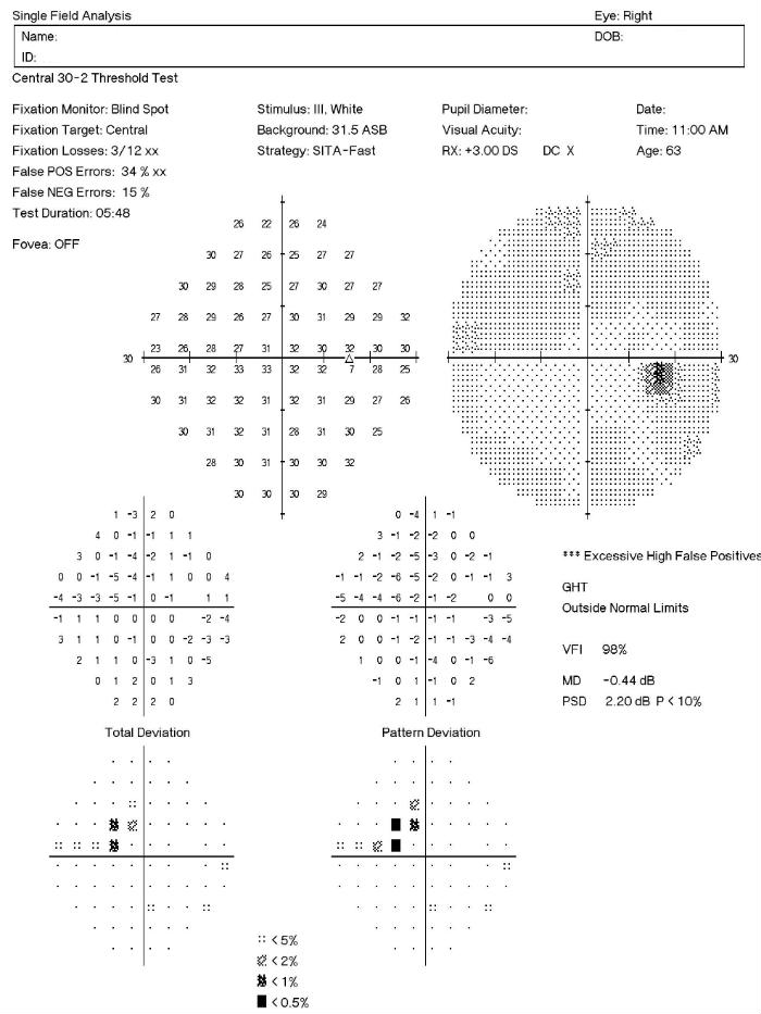 |
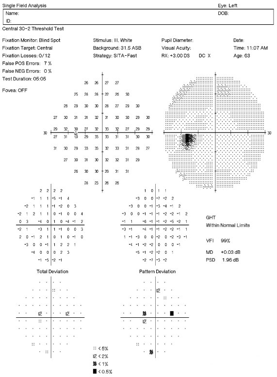
Visual Field Examination Test Results in a 64-year-old Glaucoma Suspect
Right Eye
|
| Visual Field Examination Test Results in a 64-year-old Glaucoma Suspect
|
 |
Gonioscopy
- Can show abnormal angle structures
- Abnormal pigmentation of the trabecular meshwork
- Abnormal anterior chamber anatomy
- Abnormal iris configuration
Corneal Pachymetry
- A central corneal thickness of less than 500 microns is a risk factor for glaucoma in the presence of elevated IOPs
Extended Ophthalmoscopy
- Evaluate optic disc morphology
- Document structural changes to the optic disc
Fundus Photography
- To document the progress or lack of progress of glaucoma
- To document the delivery of medical treatment
- To document the response to treatment
- To help plan a treatment program
- Fundus autofluorescence imaging can be used to detect structural abnormalities and predict functional deficits before standard color fundus photography
- Multi-spectral imaging with the Annidis RHA System can be used to detect structural abnormalities and predict functional deficits before standard color fundus photography
 |
 |
Electrodiagnostics
- Visual evoked potential testing evaluates the function of the afferent visual sensory system
- Electroretinography evaluates the function of the retinal ganglion cells
 |
|
Serial Tonometry
- Establish diurnal curve
Ultrasound Biomicroscopy (UBM) and/or Anterior Segment Imaging
- Determine iris configuration
- Evaluate anterior chamber anatomy
- Determine the mechanism of primary glaucoma
- Determine the occludability of the angle
Refraction
- Measurement of visual acuity
- Measurement of visual function
Provocative Tests for Glaucoma
- Risk assessment for angle-closure glaucomas
Open-angle glaucoma suspects are classified based on the number of risk factors present.
- Low risk — One or two risk factors
- High risk — Three or more risk factors
Differential diagnosis will depend on if the damage is observed on the optic nerve or a visual field defect is present. Below is a list of possible differential diagnosis.
Optic Nerve Head
- Congenital and hereditary anomalies of the disc
- Ischemic optic neuropathy
- Compressive lesions of the optic nerve
Visual Field Defect
- Pituitary tumors
- Ischemic optic neuropathy
- Vascular disease
- Drusen of the optic nerve
- Neurological condition
When initiating treatment, assume that the pre-treatment measurement of intraocular pressure (IOP) is the level that produced damage to the optic nerve. If the IOP remained at this level, additional damage to the optic nerve would follow.
To treat glaucoma, an IOP level is identified below which further optic nerve damage is unlikely to occur. This IOP level is the target pressure range and it is selected based on the following:
- Pre-treatment level of IOP
- The rapidity with which the damage to the optic nerve occurred, if that is known
- Patient age
- General health of the patient
Treatment should maintain the IOP at or below the target level. The status of the optic nerve, visual fields, electrodiagnostics, and the retinal nerve fiber layer are monitored over time for evidence of stability or deterioration. In the event of further damage, the target pressure range is reset to a lower level.
Pharmacologic Treatment
Select your initial medication based upon target pressure range, medical history, and ocular history. If the initial medication fails to achieve the target pressure range within one month, discontinue the initial medication and substitute another.
If the second medication fails to achieve the target pressure, begin combination therapy by using two different medications. If this strategy fails, you could add still a third class of glaucoma medications to the regimen. With three different eye drops, non-compliance becomes common and laser surgery may be a better treatment option.
Prostaglandins are the most popular class of medications for glaucoma therapy due to their excellent efficacy, safety index and tolerability. They flatten the diurnal curve significantly and achieve the greatest IOP reduction of any class of topical medication.
Classes of Glaucoma Medications
Prostaglandins
- Lumigan
- Travatan Z
- Zioptan
- Xalatan
Beta-Blockers
- Timolol
- Betagan
- Betimol
- OptiPranolol
Adrenergic Agonists
- Alphagan P 0.1% and 0.15%
- Brimonidine 0.2%
- Propine
Carbonic Anhydrase Inhibitors
- Azopt
- Trusopt
Cholinergic Agonists
- Pilocarpine
- Carbachol
- Echothiophate
Combination Glaucoma Medications
- Cosopt – carbonic anhydrase inhibitor / beta-blocker
- Cosopt PF – carbonic anhydrase inhibitor / beta-blocker
- Combigan – adrenergic agonist / beta-blocker
- Simbrinza – adrenergic agonist / carbonic anhydrase inhibitor
Changing the Treatment due to Side Effects
Prostaglandins
- Red eye
- Eye color change
- Excessive eyelash growth
- Uveitis
- Macular edema
Beta-Blockers
- Bronchospasm
- Pulmonary edema
- Heart failure
- Headache
- Weakness
- Depression
- Lethargy
- Itching
- Stinging
- Blurred vision
- Photophobia
- Loss of libido
Adrenergic Agonist
- Red eye
- Allergic follicular conjunctivitis
- Dry mouth
- Drowsiness
Carbonic Andydrase Inhibitors
- Sulfa allergy
- Red eye
- Ocular itching
- Metallic taste
- Paresthesia of fingers or toes
- Headache
- Blood dyscrasias
- Depression
- Loss of libido
- Impotence
Cholinergic Agonists
- Red eye
- Induced myopia
- Reduced vision in low illumination
- Headaches
- Lens opacities
- Iris cysts
- Increased risk of angle closure
- Increased risk of retinal detachment
- Sweating
- Salivation
- Diarrhea
- Bradycardia
- Dyspnea
Docosanoids
- Eye color change
- Stinging
- Increase in periocular eyelid pigmentation
- Increase in upper eyelid sulcus deepening
1. Ventura LM, Golubev I, Feuer W, Porciatti V. The PERG in Diabetic Glaucoma Suspects With No Evidence of Retinopathy. J Glaucoma 2010; 19(4): 243-247.
2. Tsai JC. VEP Technology for the Detection of Glaucomatous Visual Field Loss. Glaucoma Today. 2009 March. http://glaucomatoday.com/2009/03/GT0309_10.php/. Last accessed June 25, 2015.
3. Banitt M, Ventura L, Feuer W, Savatovsky E, Luna G, Shif O, Bosse B, Porciatti V. Progressive Loss of Retinal Ganglion Cell Function Precedes Structural Loss by Several Years in Glaucoma Suspects. National Institutes of Health-National Eye Institute. 7 Feb 2013. http://www.ncbi.nlm.nih.gov/pmc/articles/PMC3626526/. Last accessed June 25, 2015.
4. Malik R, Swanson W, Garway-Teath D. The ‘Structure-function’ Relationship in Glaucoma — Past Thinking and Current Concepts. Clin Experiment Ophthalmol. 2012 May-Jun;40(4):369-80. doi: 10.1111/j.1442-9071.2012.02770.x. Epub 2012 Apr 12. http://www.ncbi.nlm.nih.gov/pubmed/22339936. Last accessed June 29, 2015.
5. Bremmer F.D. Pupil Assessment in Optic Nerve Disorders. Eye (Lond). 2004 Nov;18(11):1175-81. http://www.ncbi.nlm.nih.gov/pubmed/15534603. Last accessed June 29, 2015.
6. Chew S, Cunningham W, Gamble G, Danesh-Meyer H. Retinal Nerve Fiber Layer Loss in Glaucoma Patients with a Relative Afferent Pupillary Defect. Invest Ophthalmol Vis Sci. 2010 Oct;51(10):5049-53. doi: 10.1167/iovs.09-4216. Epub 2010 May 5. http://www.ncbi.nlm.nih.gov/pubmed/20445112. Last accessed June 29, 2015.
7. Tielsch JM, Sommer A, Katz J, Royall RM, Quigley HA, Javitt J. Racial Variations in the Prevalence of Primary Open-Angle Glaucoma The Baltimore Eye Survey. JAMA. 1991 Jul 17;266(3):369-74. http://www.ncbi.nlm.nih.gov/pubmed/2056646. Last accessed June 29, 2015.
8. Rabin J, Gooch J, Ivan D. Rapid Quantification of Color Vision: The Cone Contrast Test. Invest Ophthalmol Vis Sci. 2011 Feb 9;52(2):816-20. doi: 10.1167/iovs.10-6283. http://www.ncbi.nlm.nih.gov/pubmed/21051721. Last accessed June 29, 2015.
365.01
Open angle with borderline findings, low risk
92133
Retinal nerve fiber laser scan
92083
Visual field examination
92250
Fundus photography
92225
Extended ophthalmoscopy
92226
Subsequent ophthalmoscopy
92020
Gonioscopy
95930
Visual evoked potential
92275
Electroretinography
92100
Serial tonometry
92132
Anterior segment imaging
76513
Anterior segment ultrasound
76514
Corneal pachymetry
Open angle glaucoma, low risk
Occurrence
The prevalence of open-angle glaucoma is almost 2% of the population over 40 years old.
Distribution
Glaucoma is not distributed evenly throughout the population.
- Black Americans 4 to 5 times more likely to develop glaucoma
- More women than men develop glaucoma
- Latino Americans have very high risk in advanced age
Risk Factors
- Vertically elongated cup-to-disc ratios
- Asymmetric cup-to-disc ratios
- Intraocular pressures greater than 21 mmHg
- Advancing age
- Black or Latino race
- Thin corneas in the presence of elevated IOPs
- Family history
- Medical history