

 Print | Share
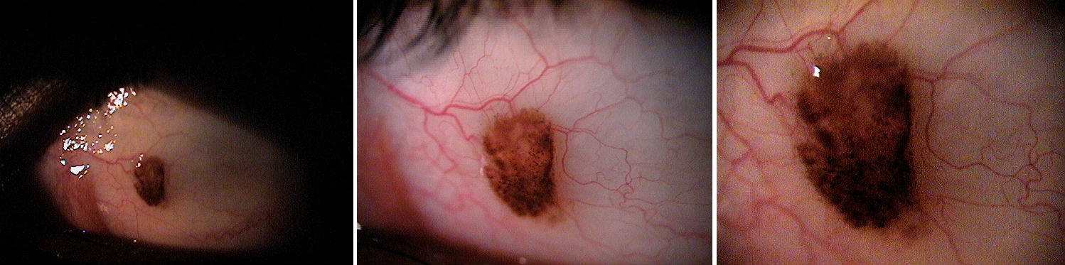
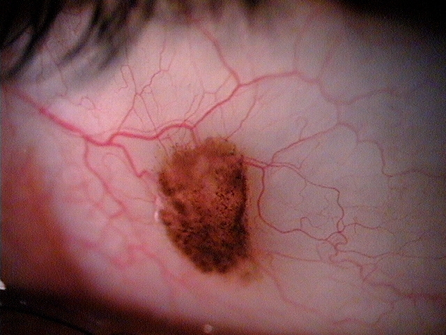
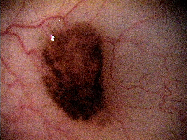
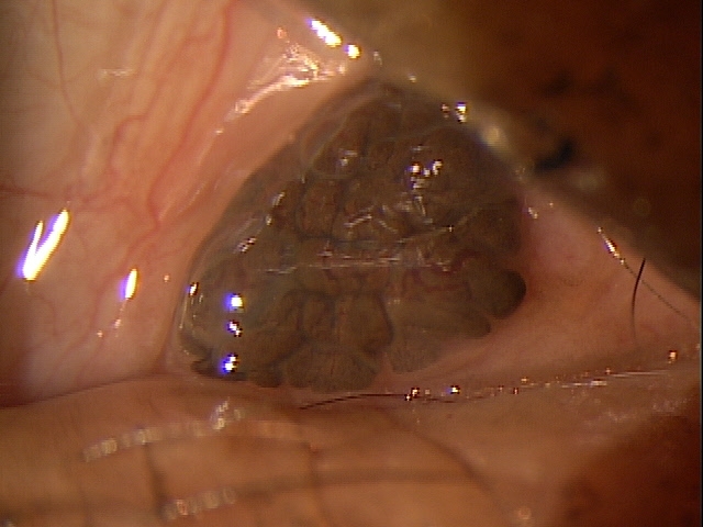
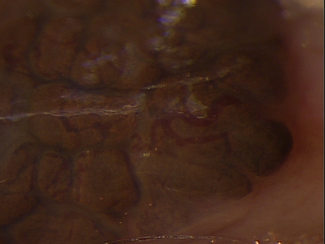
Pigmented lesion overlying the caruncle
ICD-10 Diagnosis Codes:
D31.01–Benign neoplasm of right conjunctiva
D31.02–Benign neoplasm of left conjunctiva
Title
Benign Neoplasm of Conjunctiva
Category
Benign Neoplasm Of Eye
Description
A neoplasm of the conjunctiva that is benign is a tumor which does not spread or “metastasize” to other parts of the body.
Benign lesions have several characteristics that differentiate them from malignant lesions. Some types of benign lesions include squamous papilloma, keratoacanthoma, hereditary benign intraepithelial dyskeratosis, keratotic plaque, actinic keratosis, pterygium and pingueculum.
Conjunctival tumors can be deadly to a patient, and with some malignant lesions mimicking benign tumors, physicians need to know what to look for.
 |
The most common benign neoplasm of conjunctiva is squamous papilloma, which is associated with human papillomavirus (HPV). It is marked by a pink, fibrovascular frond of the tissue, either sessile or pedunculated. Its pathology shows finger-like projections of fibrovascular tissue, usually acanthotic, with hyperkeratosis. It can be treated with surgical excision, cryotherapy or topical chemotherapy.
Keratoacanthoma lesions are inflammatory lesions that might mimic carcinoma. They are raised, fleshy, oftentimes nodular lesions; though sometimes they are gelatinous or leukoplakic that onset rapidly. Pathologically, they are brought on by hyperkeratosis and acanthosis with neutrophilic infiltrate and microabscesses. They should be treated with excision.
Keratotic plaque appears as a white conjunctival mass with keratinized, acanthotic epithelium. It appears similar to squamous cell carcinoma with leukoplakia.
Actinic keratosis appears as a frothy white lesion often located over a pingueculum or pterygium. It shows dysplastic, keratotic epithelium and also can resemble squamous cell carcinoma. It is autosomal dominant disorder with elevated fleshy plagues and shows acanthosis, dyskeratosis and inflammatory cells.
External Ocular Examination with Biomicroscopy
|
 |
|
Clinical Appearance of the Conjunctiva
|
 |
|
 |
Clinical Appearance of the Conjunctiva
|
|
 |
Clinical Appearance of the Conjunctiva
|
Neoplasms of the eye are classified as either choroidal or conjunctival, depending upon their location. A neoplasm of the conjunctiva can be classified as benign or malignant, depending on if can metastasize to other parts of the body.
- Conjunctival melanoma
- In pigmented lesions of the conjunctiva, a biopsy is contraindicated
- If the lesion turns out to be a melanoma, a biopsy eliminates the possibility of getting clear margins going forward
- Therefore, standard therapy for a rapidly growing pigmented lesion on the conjunctiva is referral to an oculoplastic surgeon for wide excision with wide margins
1. Laber D. Symptoms, treatment for eye lesions. Eyeworld. Nov 2009. http://www.eyeworld.org/article-symptoms–treatment-for-eye-lesions. Last accessed January 30, 2015.
2. Sayyad F, Karp C. Conjunctival Pigmented Lesions: Diagnosis and Management. American Academy of Ophthalmology. Sept 2013. http://www.aao.org/publications/eyenet/201309/pearls.cfm. Last accessed January 31, 2015.
3. Roque M. Conjunctival Melanoma. Medscape/EMedicine. 9 Oct 2013. http://emedicine.medscape.com/article/1191840-overview#showall. Last accessed January 31, 2015.
224.3
Benign neoplasm of conjunctiva
92285
External ocular photography