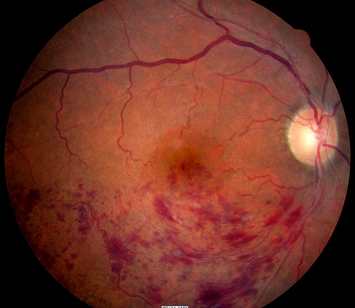
Branch retinal vein occlusion
ICD-10 Diagnosis Codes:
H34.831–Branch retinal vein occlusion, right eye
H34.832–Branch retinal vein occlusion, left eye
H34.833–Branch retinal vein occlusion, bilateral
Title
Branch Retinal Vein Occlusion
Category
Other Retinal Disorders
Description
A branch retinal vein occlusion (BRVO) occurs when one of the branches of the central retinal vein carrying blood away from the retina becomes occluded.
A branch retinal vein occlusion (BRVO) is a common intraretinal vascular event. It is caused by the occlusion of one of the branches of the central retinal vein, and is usually located distal to the central retinal vein’s bifurcation.
Although most branch vein occlusions occur in patients with systemic vascular disease, the events may occur spontaneously and without cause.
The pathogenesis of a branch retinal vein occlusion depends upon the vascular perfusion status. For example, a non-ischemic occlusion occurs without interruption of vascular profusion. Conversely, an ischemic occlusion is characterized by vascular stasis and the side effects of limited blood circulation.
- Turbulent blood flow
- Damage to the vascular endothelium
- Thrombosis formation
Structural Damage to the Eye
- Retinal ischemia
- Macular edema
Functional Damage to the Eye
- None
- Loss of visual acuity is dependent on the amount of macular involvement
The main goal of the diagnostic evaluation for a patient with BRVO is to accomplish the following:
- Differentiate between ischemic and non-ischemic occlusions
- Determine the underlying systemic cause of the occlusion
- Prescribe a treatment plan
Patient History
Patients with BRVO will present with the following clinical symptoms or signs:
- Painless, gradual to sudden vision loss
- Visual disturbances such as flashes of light or floaters
- An abnormally intraocular pressure (IOP) will help differentiate BRVO versus ocular ischemic syndrome from a carotid occlusive disease. Patients with ocular ischemic syndrome will have lower IOP than patients with BRVO
- Ischemic BRVO will have a relative afferent pupillary defect
DIAGNOSTIC TESTS
Refraction
- Measurement of visual function / visual acuity
- Visual acuity of 20/400 or worse with correction along with signs of an afferent pupillary defect and visual field constriction are signs of an ischemic BRVO
Blood Pressure Measurement
- Systemic hypertension is a risk factor for branch retinal vein occlusion
Fundus Photography
- To evaluate the optic nerve, macula and the retina
Extended Ophthalmoscopy
- To evaluate the optic nerve, macula and the retina
Visual Field Examination
- Measurement of visual function
Macular OCT Scan
- Evaluate structural changes to the retinal and choroidal blood vessels
Gonioscopy
Any patient with a history of ischemic eye disease should receive a gonioscopic examination of the anterior chamber angle to look for any of the following:
- Fibrovascular membranes
- Neovascular blood vessels
- Angle-closure
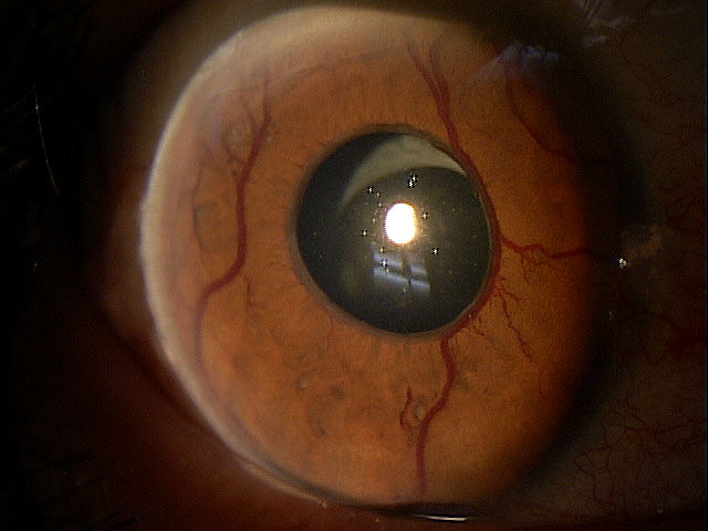 |
Iris Neovascularization
|
Systemic Evaluation
Eye doctors should consider a systemic evaluation in cases of branch retinal vein occlusion. A typical investigative protocol would include the following tests:
Blood Work
- Complete blood count (CBC)
- Westergren sedimentation rate (ESR)
- Lipid profile
- Fasting blood sugar/HbA1C
- Fasting treponemal antibody absorption test (FTA-Abs)
- Reactive plasma reagin (RPR)
- Homocysteine
- Protein S and C
- Lupus anticoagulant
- SickleDex
- Rheumatoid factor
- Purified protein derivative (PPD)
- Lyme titer
- Angiotensin converting enzyme (ACE)
- Antinuclear antibody (ANA)
Ancillary Testing
- Carotid Doppler
- Electrocardiogram (EKG)
- Chest X-ray
Retinal vein occlusions may be identified by their location in the vascular arcades:
- Superior temporal
- Inferior temporal
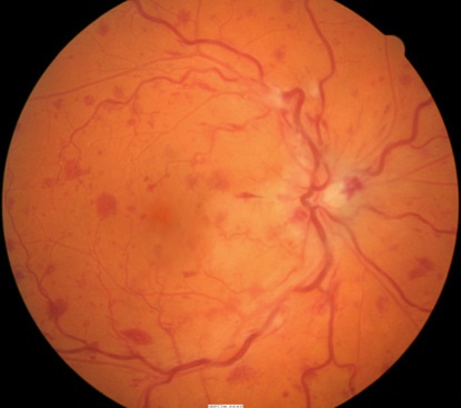 |
Nonischemic Branch Retinal Vein Occlusion
The milder form of the disease. Patients present with good vision, few retinal hemorrhages and cotton-wool spots, no relative afferent pupillary defect, and good perfusion to the retina. Nonischemic BRVO may resolve fully with good visual outcome or may progress to the ischemic type. |
|
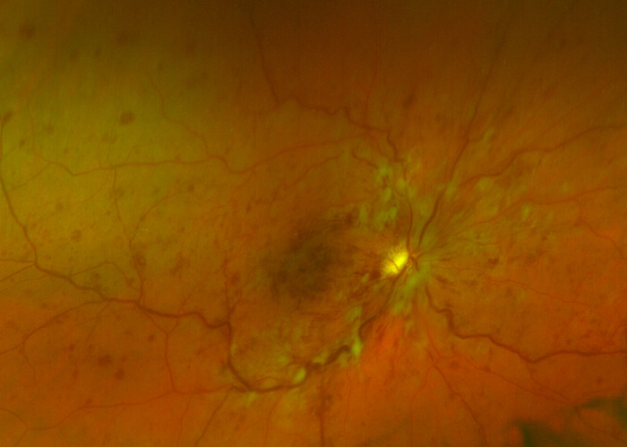 |
Ischemic Branch Retinal Vein Occlusion
The severe form of the disease. BRVO may present initially as the ischemic type, or it may progress from nonischemic. Usually, ischemic BRVO presents with severe visual loss, extensive retinal hemorrhages and cotton-wool spots, presence of relative afferent pupillary defect and poor perfusion to retina. Other complications may arise such as neovascular glaucoma and a painful blind eye. Ischemic BRVO can be better distinguished with fluorescein angiography. |
Ocular Ischemic Syndrome (Carotid Occlusive Disease)
Patient will have dilated and irregular veins, but they will not be tortuous like in CRVO. Neovascularization is present in a third of patients, but disc edema and hemorrhage are not common. Retinal hemorrhages are found in the mid-periphery of the retina. There will be a history of transient ischemic attacks, amaurosis fugax and orbital pain. Intraocular pressures is usually low.
Diabetic Retinopathy
The hemorrhages and microaneurysms are concentrated in the posterior pole and exudates are more common. It is usually bilateral.
Papilledema
There will be bilateral disc swelling with flame-shaped hemorrhages around the disc, but not into the peripheral retina. Etiology is caused from increased intracranial pressure rather from a vascular etiology
Radiation Retinopathy
There is a history of radiation therapy. Patients will have disc swelling and retinal neovascularization, and cotton wool spots.
The goals of treating a branch retinal vein occlusion include the following:
- Differentiate the occlusion from ischemic vs. non-ischemic
- Treat any clinically significant secondary macular edema
- Identify the underlying systemic cause for the occlusion
- Treat the systemic disease in cooperation with internal medicine
Non-Surgical Treatment Guidelines
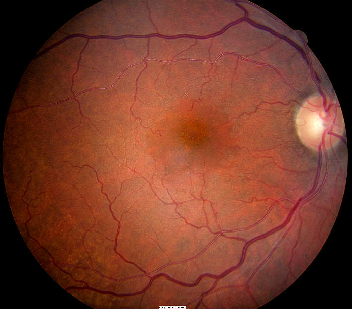
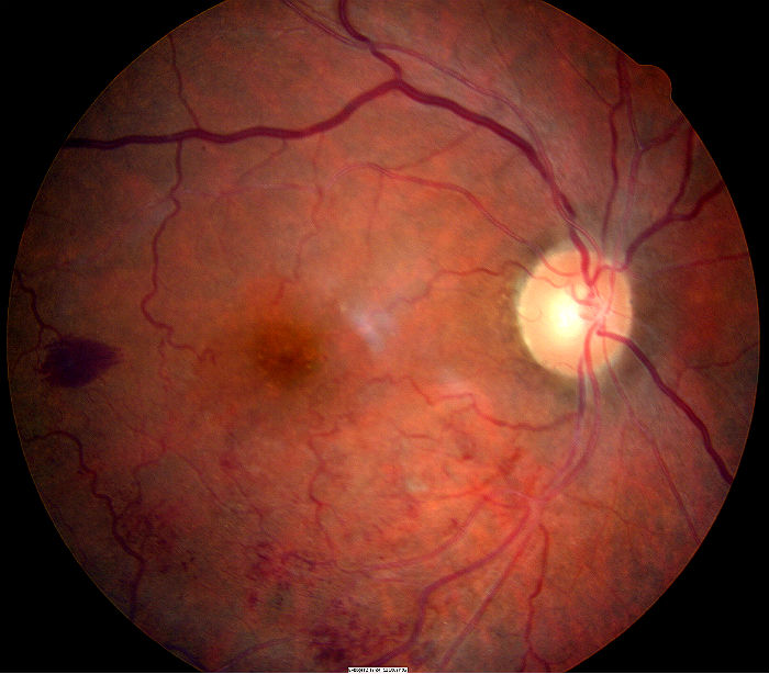
Observation for spontaneous resolution is an option in eyes with 20/40 vision or better. Even in the presence of mild macular edema, some patients may decline intravitreal anti-VEGF injections or laser surgery. In patients where observation is chosen as the initial treatment option, follow-up examinations may be performed at 3-month intervals. Non-surgical treatment involves close monitoring of the condition both on a home and clinical basis to maximize the earliest detection of further vision deterioration. This is best accomplished by home Amsler grid testing and repeat ophthlalmoscopic evaluations including OCT analysis.
The patient in the photographs below is a 95-year-old black female who declined all surgical and pharmacologic intervention.
- Mild macular edema
- Retinal hemorrhage resolved over six months duration
- Distance visual acuity was never worse than 20/40 during the course of observation
 |
 |
 |
||
| January 2013 No retinal pathology |
March 2014 Recent branch retinal vein occlusion |
September 2014 Resolving BRVO 6 months later |
Laser Therapy
Laser photocoagulation is the treatment of choice in complications associated with retinal vascular diseases (i.e. diabetic retinopathy, branch retinal vein occlusion). Panretinal photocoagulation (PRP) has been used in the treatment of neovascular complications of BRVO for a long time. However, no definite guidelines exist regarding exact indication and timing of PRP.
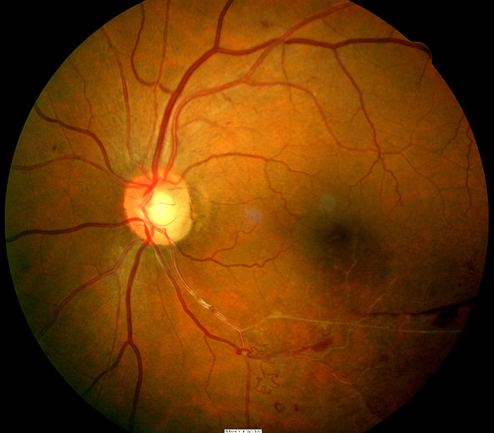
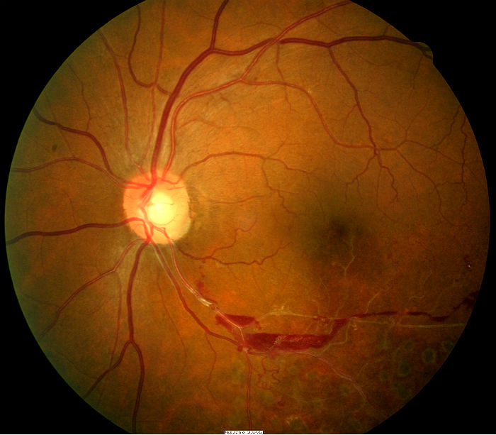
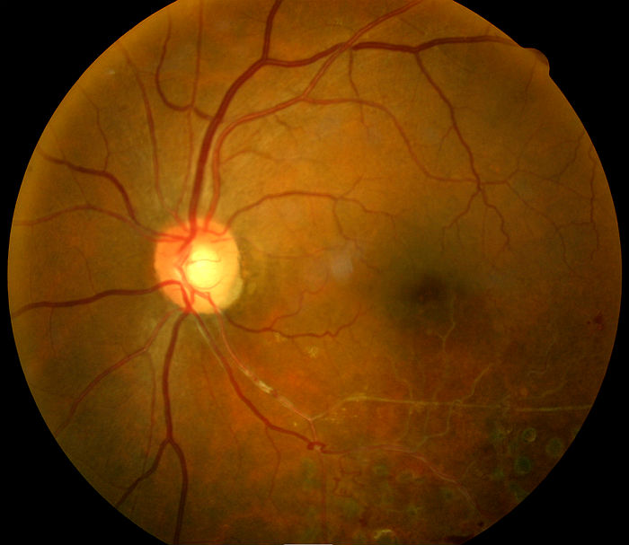
The patient in the photographs below is a 62-year-old black female with a six-month history of decreased vision in the left eye..
- Old branch retinal vein occlusion along the inferior temporal arcade
- 20/30 visual acuity
- Normal Amsler grid testing
- No macular edema on OCT scan
- Several areas of neovascularization are present
- Laser surgery recommended to prevent secondary vitreous hemorrhage
 |
 |
 |
||
| Branch retinal vein occlusion with neovascularization treated with panretinal photocoagulation today | 3 months after initial visit — retinal hemorrhage still present — 20/20 visual acuity | 10 months after initial visit — retinal hemorrhage resolved — 20/20 visual acuity |
1. Wu L. Branch Retinal Vein Occlusion. EMedicine/Medscape. http://emedicine.medscape.com/article/1223498-overview. Last accessed December 5, 2014.
2. Sowka J. Kabat A. Occlusion Conclusions. RevOptom. 14 May 2010. http://www.reviewofoptometry.com/content/d/therapeutic_review/i/1104/c/20809/. Last accessed August 17, 2014.
3. McGinnis J. Retinal Venous Occlusion. RevOptom: The 11th Annual Guide to Retinal Disease. 2014 Nov; 18-22.
4. Dinardo A, Walling P. Going Back to Gonio. RevOptom. 15 Oct 2013. http://www.reviewofoptometry.com/content/c/44408/. Last accessed December 6, 2014.
362.36
Branch retinal vein occlusion
92134
Macular OCT scan
92250
Fundus photography
92020
Gonioscopy
92083
Visual field examination
92225
Extended ophthalmoscopy
92283
Color vision examination
92275
Electroretinography
Occurrence
- Patients who are usually affected are in their fifth or sixth decade of life
Distribution
- No predilection is apparent based on sex or race
Risk Factors
- Systemic hypertension
- Other systemic vascular disease
- Alcohol consumption


 Print | Share