

 Print | Share
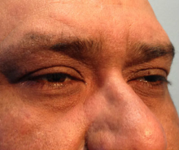
Floppy eyelid syndrome
ICD-10 Diagnosis Code:
H02.89–Other specified disorders of eyelid
Title
Floppy Eyelid Syndrome
Category
Inflammation Of Eyelids
Description
Floppy eyelid syndrome (FES) is an under-diagnosed entity characterized by chronic papillary conjunctivitis in upper palpebral conjunctiva that is poorly respondent to topical lubrication and steroids.
Floppy eyelid syndrome (FES) is a condition usually seen in overweight people. It is characterized by upper eyelids that are floppy, rubbery, and easily everted and is associated with a variable chronic papillary conjunctivitis of the upper palpebral conjunctiva.
Floppy eyelid syndrome, because its symptoms are common to other disease processes, often is not diagnosed at the onset of symptoms. Several unsuccessful trials of artificial tears, vasoconstrictors, topical steroids, nonsteroidal anti-inflammatory drops, or antibiotics may already have taken place before the correct diagnosis is made. Although FES has been reported in nonobese patients, it is seen more frequently in patients who are obese. The condition often is associated with obstructive sleep apnea.
Structural Damage to the Eye
- Dry eye
- Red eye
- Chronic irritation
- Discharge
- Corneal ulcers
- Corneal scarring
Functional Damage to the Eye
- Vision loss secondary to corneal epitheliopathy
The main goal of the diagnostic evaluation in a patient with floppy eyelid syndrome is to accomplish the following:
1. Stage the disease based on it’s clinical features
- Patient symptoms
- Eyelid appearance
- Ocular surface staining
2. Relieve ocular pain and discomfort
- Prescribe a treatment program to treat the anterior blepharitis
- Prescribe a treatment program to treat any associated dry eye syndrome
Patient History
Patients may present with any or all of the following clinical signs and symptoms:
- Unilateral or bilateral chronic eye irritation and burning
- Tearing
- Ropy, mucoid discharge (usually worse in the morning)
- Decreased vision (if there is an associated keratopathy)
- Daytime somnolence
- Morning headaches
External Ocular Examination with Biomicroscopy
- Lax upper eyelid that is easily everted when pulled superiorly toward the eyebrow
- Soft and rubbery tarsal plate that can be folded upon itself
- Laxity that can be quantified through measurement of anterior eyelid distraction
- Atrophic tarsal plate
- Stringy, mucoid conjunctival discharge
- Punctate corneal epitheliopathy and mucous strands in the tear film and fornices (possibly)
- Eyelash ptosis with loss of lash parallelism (i.e., lashes point downward toward the cornea and curve in different directions)
DIAGNOSTIC TESTS
External Ocular Photography
- Document clinical appearance of the eyelid margin
- Document clinical appearance of the inferior cornea
- Formulate a treatment plan
There is no classification system in place for floppy eyelid syndrome.
Several conditions that are similar in nature to floppy eyelid syndrome are:
- Adult blepharitis
- Adult ptosis
- Allergic conjunctivitis
- Atopic keratoconjunctivitis
- Chalazion
- Contact lens complications
- Dacryocystitis
- Demodicosis
- Dermatochalasis
- Ectropion
- Giant papillary conjunctivitis
- Hordeolum
- Keratoconus
- Ocular rosacea
- Psoriasis
- Superior limbic keratoconjunctivitis
The goals of treating floppy eyelid syndrome include the following:
- Improve patient symptoms
- Reduce chronic eyelid inflammation
- Prevent damage to the ocular surface
Surgical Treatment
- Upper and lower eyelids can be tightened at the lateral canthus by using a standard lateral tarsal strip procedure
- Horizontal shortening of the lateral upper eyelid can be achieved by performing a full-thickness resection of the lateral one fourth to one third of the eyelid margin. This can be accomplished by means of a vertical full-thickness resection up to an eyelid crease incision. Ptosis repair or blepharoplasty can be performed at the same time. The disparity in skin length can be managed with a vertical Burow triangle directed toward the brow at the lateral extent of the eyelid crease incision. A modified curvilinear back-tapered full-thickness resection with an advancement flap at the lateral upper eyelid has also been described.
- In cases with more medial laxity, horizontal shortening of the medial upper eyelid can be achieved by performing a laterally displaced pentagonal full-thickness resection in the medial one third of the eyelid, lateral to the superior punctum. Any brow ptosis, dermatochalasis, blepharoptosis, or ectropion can be repaired at the same time.
- In repairing ptosis of a lax upper eyelid, the eyelid often must be tightened to achieve the desired contour.
- Complications of surgical treatment of floppy eyelid syndrome include the following: (1) poor wound healing (2) unacceptable eyelid height or contour (3) undercorrection or overcorrection
1. Ventocilla M. Floppy Eyelid Syndrome. Medscape/EMedicine. 10 Sept 2014. http://emedicine.medscape.com/article/1212978-overview#showall. Last accessed December 14, 2014.
2. Floppy Eyelid Syndrome. RevOptom: Handbook of Ocular Disease Management. March 2004. http://www.reviewofoptometry.com/cmsdocuments/2010/4/ro0410_hndbk.pdf. Last accessed December 14, 2014.
374.89
Other disorders of eyelid
92285
External ocular photography
Distribution
- Commonly middle-aged patients (40-50 years)
- Patients 25-80 years
- Incidence slightly higher in men than women
Risk Factors
- Obesity
- Males are affected more than females
- Hypertension