

 Print | Share
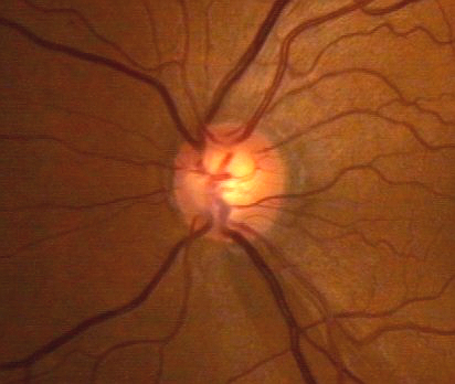
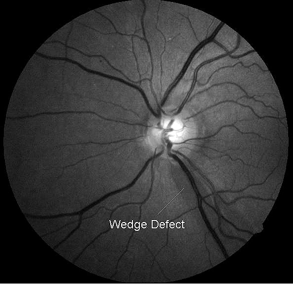
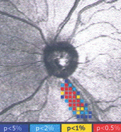
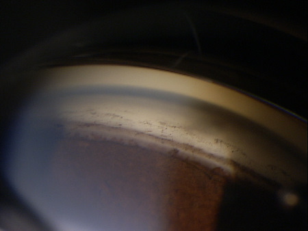
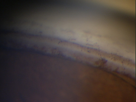
Inferior notch of the left optic disc with
associated nerve fiber layer defect.
Case Report ID: 3
Title
Normal-Tension Glaucoma; Moderate Damage
Category
Glaucoma (8)
Description
This case present the diagnosis and treatment of normal-tension open-angle glaucoma. In normal-tension glaucoma, the intraocular pressure is never measured above 21mm Hg.
Glaucoma is an optic neuropathy showing distinctive changes in optic nerve morphology without associated pallor. The term“glaucoma” refers to a group of chronic, progressive optic neuropathies that have in common characteristic morphologic changes at the optic nerve and retinal nerve fiber layer.
The glaucomas are associated with the following clinical features:
- Aqueous outflow restrictions
- Unphysiologic intraocular pressure
- Abnormal ocular perfusion
- Abnormal rate of apoptosis
- Progressive retinal ganglion cell loss
- Characteristic changes in optic nerve anatomy
Case Report
- A 60-year-old black woman presented with a chief complaint of decreased vision
- Case history described a subjective decrease in vision over the past year
- The patient had not been examined in several years and used over-the-counter reading glasses as her only visual aid
Conclusion
Glaucoma is a diagnosis of exclusion. In particular, the diagnosis of normal-tension glaucoma can be difficult. To avoid missing the diagnosis, remember that “normal” intraocular pressure relates to the statistical sense and is no guarantee that the patient does not have glaucoma.
Additionally, caution should be exercised in making the diagnosis of “normal tension” glaucoma without multiple recordings of intraocular pressure at several times during the day. Intraocular pressure can vary dramatically during the twenty-four hour daily cycle, from day to day, and when influenced by environmental factors. As thorough an understanding of a patient’s intraocular pressure is essential before beginning any treatment regimen.
History of Present Illness
- Associated Signs and Symptoms: none
- Location: vision is worse in the left eye
- Duration: past one year
- Quality: n/a
- Context: decreased vision is more noticeable when driving at night
- Severity: decrease in vision is mild
- Timing: vision seems to be getting worse over time
- Modifiers: none
Review of Systems
- The patient reported that she was in good health and taking no medications
Past, Family and Social History
- Non-contributory
Uncorrected Distance Visual Acuity
- 20/20 in the right eye
- 20/20 in the left eye
Normal Examination Findings
- Mental status
- General medical observation
- Pupils
- Basic sensorimotor examination
- External examination
- Adnexal examination
- External ocular examination with biomicroscopy
Intraocular Pressure Measurements
- 14 mm Hg in the right eye
- 18 mm Hg in the left eye
Population studies indicate that the average IOP is 15.5 mm Hg +/- 2.6 mm Hg.
Some studies suggest that there is a relationship between between the amount of intraocular pressure asymmetry and the likelihood of having glaucoma.
A difference in IOP of 0 mm Hg correlates with a 0.5% chance of having glaucoma
A difference in IOP of 3 mm Hg correlates with a 10% chance of having glaucoma
A difference in IOP of 6 mm Hg correlates with a 50% chance of having glaucoma
A difference in IOP of more than 6 mm Hg correlates almost 100% of the time with patients having glaucoma
As an aside, this patient was seen on multiple occasions with IOP readings never recorded above 19 mm Hg in either eye.
Gross Visual Fields
- Confrontation testing revealed a constriction in the superonasal quadrant of the left eye
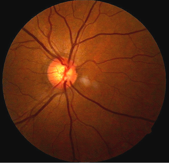
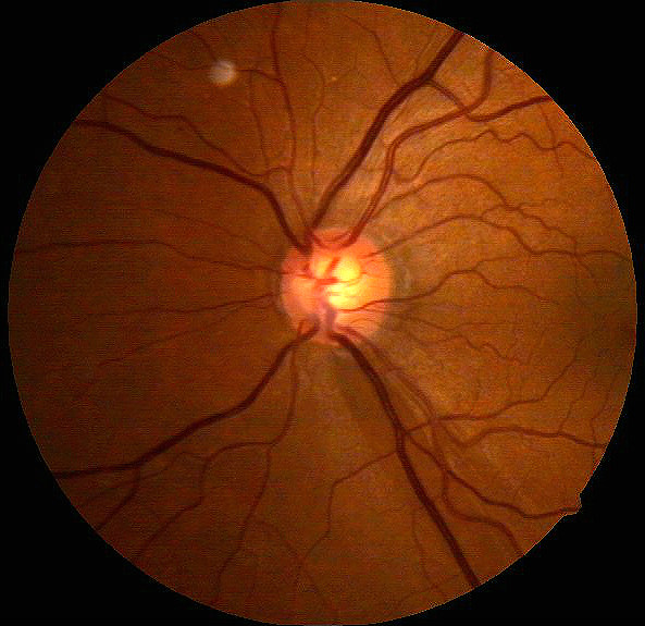
Ophthalmoscopy
- Cup-to-disc ratio = .50/.60 in the right eye
- Cup-to-disc ratio = .65/.70 in the left eye
- Vertical elongation of both optic cups
- Focal notch inferiorly on the left optic disc
- Nerve fiber layer defect in the inferior portion of the left retina
 |
 |
|
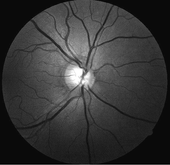 |
Retinal Nerve Fiber Layer
|
| Retinal Nerve Fiber Defect
Defects can occur in any of the following diseases:
|
 |
Glaucoma is a disease that must be classified by type to be treated properly. After a clinical diagnosis has been determined, the diagnostic process continues with a process that involves the identification and exclusion of differential diagnoses. The differential diagnosis process allows the eye doctor to distinguish between two or more diseases with similar signs and symptoms by systematically comparing their signs and symptoms.
Differential Diagnoses
The first differential diagnosis that needs to be determined in this patient is glaucoma vs. some other retinal or neurological disease. The list of possible diseases would include any pathology that produces an asymmetric loss of neuroretinal rim tissue, a corresponding retinal nerve fiber layer defect and an associated visual field defect.
The following diseases share some of the clinical signs and symptoms of glaucoma:
- Retinal detachment
- Pituitary tumors and other neurological conditions
- Ischemic optic neuropathy
- Vascular disease
- Drusen of the optic disc
- Congenital disc anomalies
Retinal detachments and drusen of the optic disc would be easily identified during routine ophthalmoscopy. Pituitary tumors producing morphologic changes in the optic nerve should be associated with characteristic bitemporal visual field defects. The patient did not have a history of vascular disease and no retinal vascular findings that would typically be associated with more advanced systemic vascular disease.
Ordering Diagnostic Tests
When additional clinical information is needed to complete the differential diagnostic process, diagnostic tests and procedures are ordered.
The performance of these tests and procedures leads to the completion of the differential diagnostic process while simultaneously initiating the treatment program. Based on the clinical diagnosis of glaucoma, the following diagnostic tests and procedures were ordered at the conclusion of the eye examination:
- Refraction
- Retinal nerve fiber layer scan
- Visual field examination
- Gonioscopy
- Corneal pachymetry
The decision to order and perform additional testing is totally based on the concept of medical necessity which can only be determined by the examining optometrist or ophthalmologist.
Refraction
- Measuring visual acuity is a method of evaluating functional vision loss
- Primary optic atrophy, especially in more severe presentations, can produce a loss in visual acuity
- 20/20 distance acuity in the right eye
- 20/25 distance acuity in the left eye
Retinal Laser Scan
- Measuring the average retinal nerve fiber layer thickness provides an overall assessment of retinal nerve fiber layer health
- The procedure can be accomplished by using the CIRRUS OCT manufactured by Carl Zeiss Meditec or other retinal scanning lasers
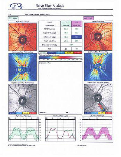 |
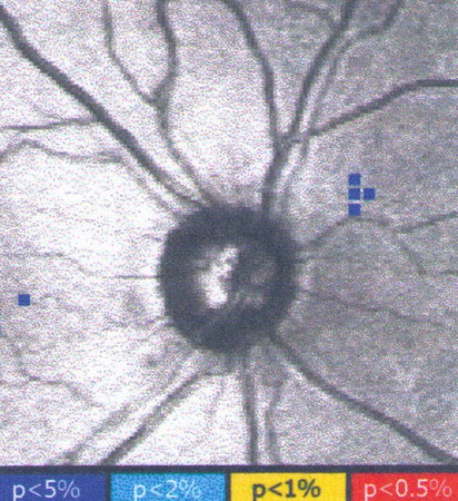
Scanning Laser Polarimetry
|
|
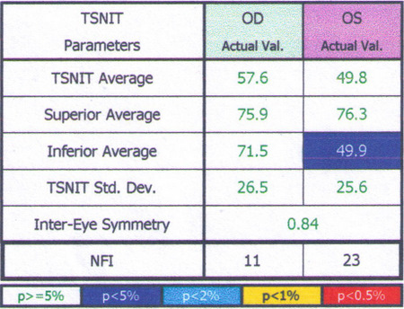 |
Inter-Eye Symmetryand theNFI Number
|
 |
 |
|
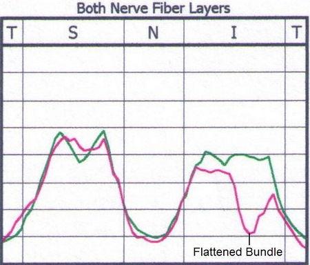 |
TSNIT Curve Profile Analysis
|
Retinal Laser Scan – Nerve Fiber Layer
- The procedure can be accomplished by the CIRRUS OCT manufactured by Carl Zeiss Meditec or other retinal scanning lasers
- OCT usually correlates with scanning laser polarimetry for retinal nerve fiber layer analysis although is considered more diagnostic as it is a direct measurement of thickness, not a calculated or extrapolated value
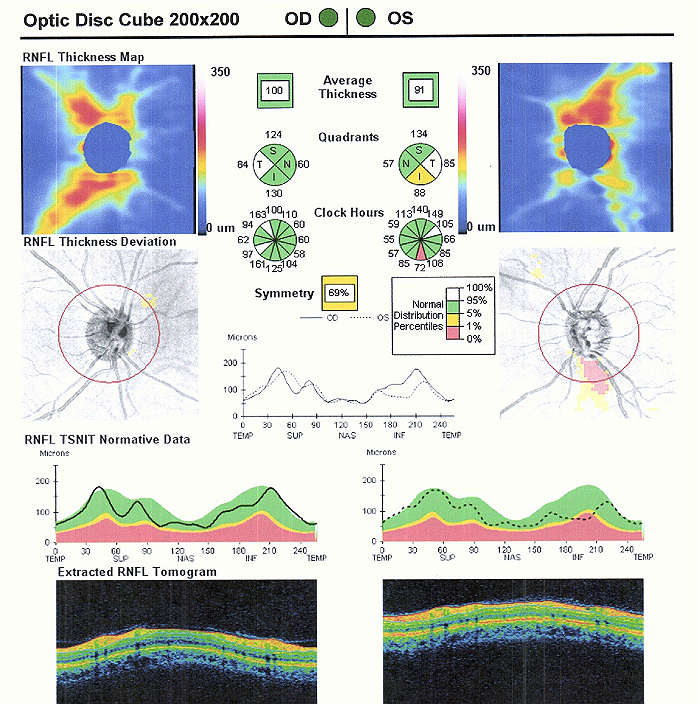 |
Right Eye
Left Eye
Both Eyes
Because glaucomatous damage to the retinal nerve fiber layer has a predilection for the inferotemporal and supertemporal regions of the optic disc, focal nerve fiber layer defects in these areas are strongly suggestive of glaucoma. |
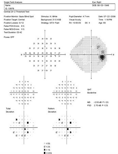
Visual Field Examination
Automated threshold perimeters measure the visual field by plotting the threshold luminescence value of the patient in various locations in the visual field. The luminescence of the light stimulus is represented by non-specific units of measurement called decibels (dB).
Glaucoma produces several changes in the visual field, and one of these changes occurs as a widespread, non-descript loss of retinal sensitivity. In Tranquair’s “Hill of Vision” concept, loss of retinal sensitivity represents a reduction in the height of the hill. The diffuse loss of retinal sensitivity should be considered highly diagnostic of glaucoma when it is asymmetric and correlates with asymmetric changes in intraocular pressure or disc appearance. In most cases, the loss of sensitivity occurs in characteristic patterns and locations (e.g., nasal step, arcuate scotoma, paracentral scotoma) that often correlate with changes in the optic nerve and/or retinal nerve fiber layer.
 |
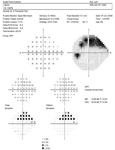 |
Automated threshold perimeters characterize specific parameters of the overall visual field status by the use of numbers called Global Indices. Two of the indices, Mean Deviation (MD) and Pattern Standard Deviation (PSD), express the raw data generated by the instrument.
A visual field defect can be classified as mild, moderate or advanced based upon an abnormal Mean Deviation.
Mild visual field defect = 0 through -5.99 dB
Moderate visual field defect = -6.00 dB through -11.99 dB
Advanced visual field defect = anything above -12.00 dB
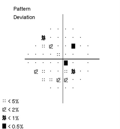
As represented by the Mean Deviation of -2.53 dB, the visual field of the right eye demonstrated a mild loss of retinal sensitivity.
Scattered paracentral scotomas were found throughout the central visual field. Paracentral scotomas are visual field defects characterized by small, isolated areas of reduced retinal sensitivity. They are typically centered just beyond the central 10 degrees of fixation but usually within 20 degrees of fixation. Paracentral scotomas occur in about 70% of patients with early glaucomatous damage to the visual system.
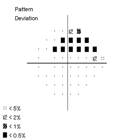
As represented by the Mean Deviation of -6.85 dB, the visual field of the left eye demonstrated a moderate loss of retinal sensitivity.
The examination revealed a constriction of the isopters to within 10 degrees of fixation. A dense arcuate scotoma was found in the superior visual field. Arcuate scotomas are visual field defects that occur when paracentral scotomas expand or coalesce with other paracentral defects. The presence of this type of defect represents moderate-to-advanced glaucomatous damage to the visual system.
The Pattern Standard Deviation is a measure of focal loss of visual field taking into account any generalized depression in the hill of vision. Many feel this index is more diagnostic than the Mean Deviation in demonstrating glaucomatous field loss while others will argue the additional “manipulation” of the data somewhat contaminates the validity of the data. The higher the PSD, the more likely the patient has glaucoma. The PSD is high for both eyes, with the left eye indicating a severe focal loss of visual field.
 |
 |
The results of the examination revealed definite glaucomatous visual field defects in each eye. The loss of retinal sensitivity should be considered highly diagnostic of glaucoma when it is asymmetric and correlates with asymmetric changes in intraocular pressure, retinal nerve fiber layer appearance or optic disc appearance. Because the visual field defect in the left eye agrees with measurable, observable structural damage to the eye, the clinical diagnosis of glaucoma is confirmed.
Gonioscopy
The anatomical configuration of the iris and angle structure is important in the pathogenesis of certain forms of glaucoma.
 |
Gonioscopy Examination Results
There is no apparent obstruction of the anterior chamber angle or outflow mechanism. |
|
 |
Goals of Performing Gonioscopic Exam of the Angle
Determinations Made After Performing Gonioscopy
|
Corneal Pachymetry
- 520 microns in the right eye
- 515 microns in the left eye
Normal central corneal thickness is approximately 550 microns. This patient’s corneal thickness profile is rated as thin.
The issue of thin corneas being a risk factor for glaucoma is currently being debated in the ophthalmic community. Recent studies suggest that an increased risk of developing glaucoma is related to abnormal cornea rigidity factors which does correspond to to corneal thickness measurements. Also, the studies showed thin central corneal thickness to be a risk factor for developing glaucoma only in patients with ocular hypertension.
Most consider corneal thickness an interesting finding that may be associated with some biomechanical risk factor in the optic nerve but otherwise not a very significant clinical finding. One should keep in mind that corneal thickness can affect the accuracy of most applanation tonometry readings and this can play a role in the classification of the glaucoma.
All of the diagnostic test results confirmed the initial diagnosis of normal-tension open-angle glaucoma, although this diagnosis may change with additional trending of intraocular pressure. Additional diagnostic tests for glaucoma such as serial tonometry, provocative glaucoma testing, or extended ophthalmoscopy were not considered medically necessary on this patient’s initial visit. Photographic documentation of the optic nerve would be an additional medically necessary test that could be obtained at any follow up evaluation.
According to Current Procedural Terminology, when eye doctors perform ophthalmological examinations, the complexity of medical decision-making is not separated from the examining techniques used. As a guideline to assist eye doctors in enhancing their medical decision-making skills, consider that the complexity of medical decision-making involves three components.
The first component concerns the number of possible diagnoses and treatment options that must be considered. Modern glaucoma diagnosis involves the determination of structural damage to the eye and/or functional loss to the visual system. Once these assessments are made, the glaucoma must be classified by type.
The second component concerns the amount and complexity of medical records and diagnostic tests that have to be obtained, reviewed and analyzed. In addition to an eye examination, this visit required the review and analysis of a subjective refraction, a retinal laser scan, a threshold visual field examination, a corneal pachymetry measurement, and a gonioscopic examination of the anterior chamber.
Third, the complexity of medical decision-making is affected by the risk of significant complications and/or morbidity associated with advanced glaucoma and the risks involved in any treatment options. This patient’s glaucoma was classified as a significant problem where the risk of total vision loss without treatment was possible. In addition, because the treatment plan would involve chronic pharmaceutical treatment with the potential for ocular and systemic side effects, the pharmacology complications had to be considered when deciding on the initial treatment options.
Treatment Guidelines
To begin the treament of glaucoma, intraocular pressure must be lowered and an initial target pressure range must be established. A general guideline is to base the initial pressure range on the category of damage to the visual system.
- Mild damage needs a 20-30% reduction in pre-treatment intraocular pressure as the initial target pressure range
- Moderate damage needs a 30-40% reduction in pre-treatment intraocular pressure as the inital target presure range
- Advanced damage needs a 40-50% reduction in pre-treatment intraocular pressure as the initial target pressure range
Doctors should always remember that the goal of glaucoma therapy is to prevent additional or future vision loss, not achieve some arbitrary target therapy range.
Due to the moderate glaucomatous damage in the left eye, a 30-40% reduction in the pre-treatment intraocular pressure was selected as the initial target pressure range.
Treatment Program
Modern glaucoma therapy generally utilizes prostaglandins as the primary therapy. The first goal of treating glaucoma is to achieve the lowest possible intraocular pressure on a single agent and prostaglandins generally have the greatest efficacy of all glaucoma medications. Monotherapy with prostaglandins improves patient compliance, may decrease costs, and improves the safety profile and tolerability of using topical medications. These drugs reduce intraocular pressure by increasing uveoscleral outflow. Intraocular pressure reduction averages almost 30% with some patients showing even greater response but a small percentage showing minimal to no response.
The most common side effect of prostaglandin therapy is conjunctival hyperemia. The redness typically diminishes over time but may be chronic. Another possible ocular side effect of prostaglandins includes changes in iris color after several months of use. In these patients, the drug produces an increase in the amount of iris stromal pigmentation. Additionally, eyelashes can lengthen, thicken, and darken – a side effect known as hypertrichosis.
One potential serious ocular side effect is a 3% increased risk of developing cystoid macular edema. A history of uveitis is a relative contraindication, as prostaglandins produce an increased risk of reactivating the inflammation. The systemic side effects of topical prostaglandins are minimal.
Avoiding medicolegal liability if complications arise is an important part of modern ophthalmic practice. This avoidance is best accomplished by making sure that you have obtained the patient’s informed consent to treat their glaucoma. Once you have made the patient fully aware that using topical glaucoma medicine involves the risks of side effects, you can proceed with the treatment plan.
After obtaining the patient’s informed consent to treat her glaucoma, Travatan Z ophthalmic solution was prescribed 1 drop at bedtime in both eyes.
A target pressure range of 10-12 mm Hg was established for each eye.
Next visit scheduled for 3 weeks.
Discussion
This case demonstrates several key aspects of glaucoma diagnosis and treatment. First, despite the moderate damage to the left eye, there was no ocular emergency and no need for a referral to a glaucoma sub-specialist at this time. Despite the glaucoma, this patient had been functioning with no alteration of life style for an indeterminable but presumably long period of time. Another few days or weeks without treatment would probably not change the course of the disease.
Second, this patient’s clinical history supports the modern understanding of the pathophysiology of glaucoma. We know that glaucoma is an optic neuropathy. Structural damage to the retinal nerve fiber layer is usually the first milestone in the natural history of glaucoma. The loss of retinal nerve fibers is closely followed by changes in the appearance of the optic disc. Functional loss, which is documented by abnormal visual field testing, abnormal visual evoked potential testing or loss of visual acuity, usually occurs later in the natural history of the disease.
On initial examination, the damage to the optic disc and the loss of visual field was moderate in the left eye. The right eye, while having no measurable fallout of the retinal nerve fiber layer, had a large optic cup and definite glaucomatous visual field defects.
Baltimore Eye Study
This patient is a reminder that the findings from the Baltimore Eye Study are still true. Although this patient was initially examined in 2008, some of the problems in diagnosing glaucoma that were revealed by the study years ago are still present.
One of the findings from the Baltimore Eye Study is that glaucoma can be hard to diagnose. The study revealed that 50% of all people found to have glaucoma during the study had seen an eye doctor within the past year and were unaware that they had glaucoma. This failure to diagnose glaucoma is most explained by a function of concentrating on intraocular pressure as a diagnostic indicator as opposed to the appearance of the optic disc.
Early Manifest Glaucoma Trial
Regarding intraocular pressures as a diagnostic indicator, the Early Manifest Glaucoma Trial demonstrated that there is considerable variability of the intraocular pressures in patients with glaucoma. The study also determined that 20-35% of glaucoma patients do not have elevated intraocular pressures. In this group of patients, the intraocular pressure will consistently measure below 21 mm Hg. In addition, the study demonstrated that 50% of patients with glaucoma – even if they had elevated intraocular pressures most of the time – had screening intraocular pressures below 22 mm Hg.
We must always be mindful that the glaucomas are a family of diseases characterized by optic neuropathy – not elevated intraocular pressure.
Based on the patient history, the nature of the presenting problem, and my own clinical judgement this patient needed an evaluation of the complete visual system.
- Perform the eye examination that is medically necessary
- Provide the diagnostic tests or services that are medically necessary
- Properly document the services provided
- Code from the documentation
- Report the services to the payor
Physicians Quality Reporting System
PQRS Measure 12: Primary Open-Angle Glaucoma: Optic Nerve Evaluation – CPT code 2027F
This measure applies to patients 18 years and older diagnosed with primary open-angle glaucoma. CPT code 2027F is reported when these patients receive an optic nerve examination. The measure should be reported on the date of the examination and on all subsequent eye examinations during the next 12 months – even if an optic nerve examination was not performed on the follow-up examinations. Remember that you may be required to report this measure more than once since the reporting period covers 12 months.
PQRS Measure 130: Current Medicatons with Name, Dosage, Frequency and Route Documented – CPT code G8427
This measure applies to patients 18 years and older. The measure should be reported on the day of the examination and can be used with any diagnosis code.
PQRS Measure 226: Patient screened for tobacco use and identified as a non-user of tobacco – CPT code 1036F
This measure applies to patients 18 years and older. The measure should be reported on the day of the examination and can be used with any diagnosis code.
Multiple Procedure Payment Reduction
Effective January 1, 2013, there is a small reduction in payment from Medicare if certain multiple procedures are billed on the same day. The fee for the technical component of the diagnostic test for the second and subsequent tests will be reduced by 20%. The second diagnostic test and subsequent tests should be reported with a -51 modifier. Professional services such as gonioscopy, extended ophthalmoscopy, serial tonometry and provocative glaucoma testing are excluded from this policy. Visual evoked potential testing is excluded from this policy.
Modifier 51
This modifier is used to identify the secondary procedure or when multiple procedures are performed on the same day by the same provider. List the major primary procedure first and append the modifier to the subsequent procedures. The primary procedure is the one with the highest dollar value.
| Diagnosis Code | Procedure Code | Modifier | Quantity | Payor | Amount Allowed |
| H40.1232 - Low-tension glaucoma, bilateral, moderate stage | 92014 - Medical eye examination | 1 | Medicare | 128.34 | |
| H40.1232 - Low-tension glaucoma, bilateral, moderate stage | 92083 - Visual field examination | 1 | Medicare | 65.21 | |
| H40.1232 - Low-tension glaucoma, bilateral, moderate stage | 92133 - Retinal laser scan | 51 | 1 | Medicare | 44.69 |
| H40.1232 - Low-tension glaucoma, bilateral, moderate stage | 92020 - Gonioscopy | 1 | Medicare | 26.96 | |
| H40.1232 - Low-tension glaucoma, bilateral, moderate stage | 76514 - Corneal pachymetry | 51 | 1 | Medicare | 14.92 |
| H40.1232 - Low-tension glaucoma, bilateral, moderate stage | 2027F - Optic nerve evaluation | 1 | Medicare | 0.00 | |
| H40.1232 - Low-tension glaucoma, bilateral, moderate stage | G8427 - Medications documented | Medicare | 0.00 | ||
| H40.1232 - Low-tension glaucoma, bilateral, moderate stage | 1036F - Patient screened for tobacco use | Medicare | 0.00 | ||
| Total | $280.12 |
H40.1232
Low-tension glaucoma, bilateral,
moderate stage
92015
Refraction
92020
Gonioscopy
92083
Visual field examination
92133
Retinal laser scan
92250
Fundus photography
95930
Visual evoked potential
92275
Electroretinography
92225
Extended ophthalmoscopy
76514
Corneal pachymetry