

 Print | Share
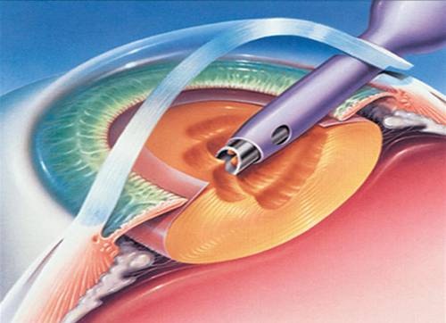
Modern cataract surgery using phaco probe
Case Report ID: 43
Title
Cataract Surgery Co-Management
Category
Peri-Operative Management Of Ocular Surgery (4)
Description
This case describes optometric co-management of cataract surgery, the management of a surgical complication (postoperative corneal edema), and the supply of prosthetic eyewear.
Cataract surgery is a common out-patient surgical procedure and complications at some level are common.
- Most complications are simply the result of inflammation from tissue manipulation and are minimal problems
- Postoperative corneal edema is one of the more common complications of cataract surgery and it is usually mild, transient and has little-to-no impact on vision
Case Report
- A 67-year-old black man presents to the office three weeks following a reported uneventful cataract surgery on the right eye
- The patient reported moderate ocular discomfort in the right eye
- The patient had already seen the operating surgeon on the one-day and one-week postoperative visit and was returning to me for my portion of the global postoperative period
General Signs and Symptoms Patient May Present at One-Day Followup
- Reduced visual acuity
- Residual dilation
- Subconjunctival hemorrhage
- Significant corneal edema
- Cells and flare in the anterior chamber
- Floaters
- Corneal keratitis and/or abrasion
- Elevated intraocular pressure
In addition, patients may also experience:
- Reduced vision
- Foreign body sensation
- Flashes and floaters
- Loss of peripheral vision
- Itching
- Burning
- Tearing
- Light sensitivity
- Grittiness
Conclusion
Co-managing optometrists must be familiar with the common complications of cataract surgery and how to properly diagnose and treat them.
History of Present Illness
- Associated Signs and Symptoms: decreased or “foggy” vision
- Location: vision is worse in the right eye
- Duration: since cataract surgery one week prior
- Quality: n/a
- Context: vision was poor immediately after surgery and has not improved
- Severity: mild
- Timing: stable
- Modifiers: none
Review of Systems
The patients medical history was unremarkable. He was using all his postoperative medications in his right eye as prescribed:
- Omnipred t.i.d.
- Nevanac t.i.d.
- Moxeza t.i.d.
Past, Family and Social History
Non-contributory.
Uncorrected Distance Visual Acuity
- 20/30 in the right eye
- 20/25 in the left eye
Normal Examination Findings
- Mental status
- General medical observation
- Pupils
- Adnexal examination
- Ophthalmoscopy
Not Performed on Today’s Examination
- Basic sensorimotor examination
- Gross visual fields
- External examination
Intraocular Pressure Measurements
- 16 mm Hg in the right eye
- 14 mm Hg in the left eye
External Ocular Examination with Biomicroscopy
- Focal area of edema in the corneal periphery
- Edematous area is characterized by a loss of stromal transparency
- Obvious thickening of the cornea highlighted by fluorescein staining
- Epithelial blisters and corneal bullae are present in the center of the lesion
- Loss of corneal luster over the area of corneal edema
 |
Clinical Appearance of the Cornea
|
|
 |
Clinical Appearance of the Cornea
|
|
 |
Clinical Appearance of the Cornea
|
|
 |
Clinical Appearance of the Cornea
|
|
 |
Clinical Appearance of the Cornea
|
Clinical Diagnosis
The clinical diagnosis is a determination based on the knowledge obtained from the patient’s medical history and from the results of the eye examination alone, without the benefit of diagnostic tests or procedures.
The patient’s clinical diagnosis is postoperative corneal edema secondary to cataract surgery based on the following clinical findings:
- Recent history of cataract surgery in the right eye
- Unexpected decreased visual acuity in right eye
- Ocular discomfort in the right eye
- Presence of bullous keratopathy on the right cornea
Treatment Plan
To gather the information required to properly counsel a patient regarding postoperative corneal edema, a diagnostic and treatment program is initiated.
- Determination of different types of diagnoses
- Selection of one or more treatment options
The diagnosis of postoperative corneal edema following cataract surgery with reduction in functional vision is relatively straightforward. There are multiple potential etiologies for chronic postoperative corneal edema. These include:
1. Primary,excessive inflammatory response to the surgery
2. Elevated postoperative intraocular pressure
3. Pre-existing corneal endothelial disease
- Fuchs’ endothelial dystrtophy
- Corneal guttata
- Abnormal rate of polymegethism
- Presence of pleomorphism
- Abnormal reduction in endothelial cell density
4. Iatrogenic corneal endotheliopathy
- Direct surgical trauma
- Endothelium touched by instruments
- Endothelium touched by IOL
- Physical damage from irrigating solutions
- Physical damage from ultrasound vibrations
5. Toxic syndromes
6. Intraocular lens syndrome
7. Inflammation from retained lens fragments
8. Detachment of Descemet’s membrane
9. Chemical injury from improperly prepared or expired solutions or from compounds retained on instrumentation
Risk factors for iatrogenic corneal endotheliopathy include preexisting endothelial disease, diabetes, glaucoma, shallow anterior chamber, and previous intraocular surgery. Every effort should be made to determine an exact etiology as treatment options will vary depending upon the cause. If a doctor other than the surgeon is performing the postoperative evaluation, the surgeon should communicate any peri-operative complications that could result in chronic postoperative corneal edema.
Ordering Diagnostic Tests
The following diagnostic tests can help determine the degree of functional vision limitation that is related to postoperative corneal edema and provide information necessary to properly develop a treatment plan and counsel the patient.
- Refraction
- Specular endothelial microscopy
- Corneal topography
- Corneal pachymetry
The decision to order and perform additional testing is totally based on the concept of medical necessity which can only be determined by the examining optometrist or ophthalmologist.
Refraction
- Measuring visual acuity is a method of evaluating functional vision loss
- Corneal edema can produce a loss of visual acuity as well as loss of quality of vision
- Mild stromal edema does not usually cause severe visual loss
- Focal areas of stromal edema located in the corneal periphery do not usually cause severe vision loss
- No improvement in vision could be obtained after subjective refraction
In many cases, the clinical appearance of the cornea is enough evidence to explain the decreased vision. In these cases, a subjective refraction is unnecessary. Pinhole testing or an automated refractor reading can help make this decision.
Specular Microscopy
Specular microscopy can both confirm the nature of the edema and quantify the degree of endothelial damage. When compared to the non-operated eye, it can also be helpful in confirming whether the cornea was likely at risk or not (although this is likely to be known from a comprehensive preoperative evaluation).
 |
Postoperative Specular Microscopy
|
Corneal Topography
Computerized corneal topography is an examination technique that allows you to analyze the physical features and shape of the anterior cornea. It combines the traditional principle of keratoscopy with the data analysis that is available from modern computers. Important functions of corneal topography include the following:
- Earlier and more accurate diagnosis of corneal disease
- Assist the doctor in determining the appropriate treatment plan
- Preoperative surgical risk assessment
- Postoperative surgical management
 |
Postoperative Corneal Topography
|
All of the diagnostic test results produced confirmed the clinical diagnosis of corneal edema. Because the patient had no evidence of corneal edema prior to the surgery, the induced edema would be classified as secondary corneal edema.
The diagnosis of surgically-induced corneal edema involves medical decision-making of low complexity. In addition to evaluating the elements of the eye in a normal postoperative evaluation, this visit required the review and analysis of a subjective refraction, a specular microscopy evaluation, and a corneal topography evaluation.
The complexity of medical decision-making is affected by the risk of significant complications and/or morbidity associated with iatrogenic corneal edema and the risks involved in any treatment options. This patient’s condition was classified as a moderate problem where the risk of significant, irreversible vision loss without treatment was low. However, medical decision-making is complicated by the wide range of treatment options available.
Treatment Guidelines
Iatrogenic corneal edema presents with a wide range of treatment options that can range from no treatment to multiple, concurrent treatments. If the edema is mild and not causing significant reduction in functional vision, no direct other than monitoring the condition is warranted. Beyond no treatment, the degree of intervention is dependent on the severity of the edema and the severity of vision impairment.
Many authorities initiate additional anti-inflammatory medications or increase dosage beyond the normal postoperative dosage of topical steroids. While inflammation is a common component of the postoperative stress on the endothelium leading to exaggerated corneal edema, unless it is a significant component, increasing steroid dosage should have no impact on the condition. Another caution in increasing the steroid dosage is the potential for increased intraocular pressure which will often add to the edema process or at a minimum slow down resolution.
If the intraocular pressure is either elevated or normally over 15-16 mm Hg, reducing the intraocular pressure can reduce the pressure gradient against the endothelium and significantly aid in reducing stromal edema. Medications that target reduction of aqueous production are preferred.
If the edema has spread through the cornea to include the epithelium, topical hyperosmotics may be of some assistance in reducing edema. Hyperosmotics will have a mild effect on stromal edema. If prescribed, hyperosmotic ointments produce less irritation and significantly greater contact time. Copius use of lubricants and application of a bandage contact lens can also help manage the edema when there is epithelial involvement.
Treatment Program
The patient was counseled regarding the nature of her disease and the treatment program explained to him. Topical steroid dosing was increased to q2h until the next progress evaluation. A progress evaluation was scheduled for 72 hours.
In addition, because the patient was now pseudophakic and had Traditional Medicare coverage, he was eligible for his prosthetic eyewear benefit administered by one of Medicare’s Durable Medical Goods Carriers.
Based on the patient history, the nature of the presenting problem, and my own clinical judgement this patient did not need an evaluation of the complete visual system.
- Perform the postoperative examination that is medically necessary
- Provide the diagnostic tests or services that are medically necessary
- Properly document the services provided
- Code from the documentation
- Report the services to the payor
Multiple Procedure Payment Reduction
Effective January 1, 2013, there is a small reduction in payment from Medicare if certain multiple procedures are billed on the same day. The fee for the technical component of the diagnostic test for the second and subsequent tests will be reduced by 20%. The second diagnostic test and subsequent tests should be reported with a -51 modifier. Professional services such as gonioscopy, extended ophthalmoscopy and provocative glaucoma testing are excluded from this policy. Visual evoked potential testing is excluded from this policy.
Modifier 51
This modifier is used to identify the secondary procedure or when multiple procedures are performed on the same day by the same provider. List the major primary procedure first and append the modifier to the subsequent procedure. The primary procedure is the one with the highest dollar value.
Modifier 55
This modifier is used when another eye doctor co-manages the patient during the global postoperative period.
Site Modifiers
Use RT for the right eye and LT for the left eye.
CATARACT SURGERY CO-MANAGEMENT — FIRST EYE
| Diagnosis Code | Procedure Code | Modifier | Quantity | Payor | Amount Allowed |
| H25.11 - Age-related nuclear cataract, right eye | 66984 - Cataract surgery | 55, RT | 90 | Medicare | 162.00 |
| H18.231 - Secondary corneal edema, right eye | 92286 - Specular microscopy | 1 | Medicare | 39.06 | |
| H52.211 - Irregular astigmatism, right eye | 92025 - Corneal topography | 51 | 1 | Medicare | 35.08 |
| Total | $225.11 |
POSTOPERATIVE EYEGLASSES — FIRST EYE
| Diagnosis Code | Procedure Code | Modifier | Quantity | Payor | Amount Allowed |
| Z96.1 - Presence of intraocular lens | V2020 - Ophthalmic frame | RT | 1 | Medicare | 63.25 |
| Z96.1 - Presence of intraocular lens | V2201 - Flat-top 28 mm bifocal lens | RT | 1 | Medicare | 117.16 |
| Z96.1 - Presence of intraocular lens | V2219 - Flat-top 35 mm add on | RT | 1 | Medicare | 98.00 |
| Z96.1 - Presence of intraocular lens | V2755 - Ultraviolet filter | RT | 1 | Medicare | 41.62 |
| Total | $320.03 |
CATARACT SURGERY CO-MANAGEMENT — SECOND EYE
| Diagnosis Code | Procedure Code | Modifier | Quantity | Payor | Amount Allowed |
| H25.12 - Age-related nuclear cataract, left eye | 66984 - Cataract surgery | 55, LT, 79 | 90 | Medicare | 162.00 |
| Total | $162.00 |
POSTOPERATIVE EYEGLASSES — SECOND EYE
| Diagnosis Code | Procedure Code | Modifier | Quantity | Payor | Amount Allowed |
| Z96.1 - Presence of intraocular lens | V2020 - Ophthalmic frame | LT | 1 | Medicare | 63.25 |
| Z96.1 - Presence of intraocular lens | V2201 - Flat-top 28 mm bifocal lens | LT | 1 | Medicare | 117.16 |
| Z96.1 - Presence of intraocular lens | V2219 - Flat-top 35 mm add on | LT | 1 | Medicare | 98.00 |
| Z96.1 - Presence of intraocular lens | V2755 - Ultraviolet filter | LT | 1 | Medicare | 41.62 |
| Total | $320.03 |
Medical Necessity
For patients who are pseudophakic single vision lenses, bifocal lenses, or trifocal lenses in frames are a covered benefit. Because coverage of refractive lenses is based upon the Prosthetic Device benefit category, there is no coverage for frames or lens add-on codes unless there is a covered lens(es). Frames provided without a covered lens(es) will be denied as noncovered.
After each cataract surgery with insertion of an intraocular lens (ICD-9 V43.1), coverage is limited to one pair of eyeglasses or contact lenses. Replacement frames, eyeglass lenses and contact lenses are noncovered. If a beneficiary has a cataract extraction with IOL insertion in one eye, subsequently has a cataract extraction with IOL insertion in the other eye, and does not receive eyeglasses or contact lenses between the two surgical procedures, Medicare covers only one pair of eyeglasses or contact lenses after the second surgery. If a beneficiary has a pair of eyeglasses, has a cataract extraction with IOL insertion, and receives only new lenses but not new frames after the surgery, the benefit would not cover new frames at a later date (unless it follows subsequent cataract extraction in the other eye).
- Tinted lenses (V2745), including photochromatic lenses (V2744), used as sunglasses, which are prescribed in addition to regular prosthetic lenses to a pseudophakic patient, will be denied as noncovered.
- Scratch resistant coating (V2760), mirror coating (V2761), polarization (V2762), deluxe lens feature (V2702) and progressive lenses (V2781) will be denied as noncovered.
- Use of polycarbonate or similar material (V2784) or high index glass or plastic (V2782, V2783) for indications such as light weight or thinness will be denied as noncovered deluxe feature.
- Specialty occupational multifocal lenses (V2786) will be denied as noncovered.
- Only standard frames (V2020) are covered. Additional charges for deluxe frames (V2025) will be denied as noncovered.
- Eyeglass cases (V2756) will be denied as noncovered.
Medicare Fee For Providing Eyeglasses After Cataract Surgery
All suppliers of DME-POS, including eyeglasses and contact lenses for postoperative cataract patients, are subject to a fee. If the optometrist/ophthalmologist is enrolled in Medicare (participating or non-participating) they may write prescriptions for their patients, but they are not registered as a supplier of DME-POS. The patient will not receive a benefit unless the supplier is DME-POS-credentialed. If the optometrist/ophthalmologist is neither a supplier nor enrolled with Medicare, the patients will not be reimbursed for their eyeglasses or contact lenses based on the doctor’s prescription or those supplied by them.
In addition, optometrists/ophthalmologists are exempt from the surety bond requirement as long as they do not fill prescriptions from outside the practice. If a new patient appears with an outside prescription from a different doctor, it is recommended to create a new record for the patient in your practice, complete with demographic information, performing visual acuities, and possibly an automated refraction.
Documentation Requirements
- An order for the lens(es) and related features must be signed and dated by the treating physician [optometrist], kept on file by the supplier, and made available upon request.
- If the ordering doctor is also the supplier, the prescription is an integral part of the patient’s record.
- The ICD-9 code that justifies the need for these items must be included on the claim.
Coding Guidelines
Deluxe lens features (V2702) include services and features such as lens edge treatments and lens drilling.
Photocromatic lenses (V2744) are those in which the degree of tint changes in response to changes in ambient light. Code V2744 is used for any type of photochromatic lens, wither glass or plastic.
Code V2745 is used for any type or color of lens tint, excluding photochromatic lenses.
Code V2755 must be used only if a UV coating is applied to a lens and not as an add-on code for the UV protection inherent in the lens material.
Anti-relective coating (V2750) is a clear lens treatment used to decrease glare and internal/external reflections.
Mirror coatings (V2761) are colored, highly reflective lens treatments.
Progressive lens (V2781) is a multifocal lens that gradually changes in lens power from the top to the bottom of the lens, eliminating the line(s) that would otherwise be seen in a bifocal or trifocal lens.
Code V2784 is an add-on used for lenses made of impact-resistant material such as polycarbonate or Trivex. Codes V2782 and V2783 (high index) must not be billed in addition to code V2784.
Codes V2100 – V2114, V2199 – V2214, V2399 – V2499, V2700, and V2770 describe specific eyeglass lenses. Only one of these codes may be billed for each lens provided. These codes include both aspheric and nonaspheric lenses.
Codes V2115, V2118, V2121, V2215, V2218 – V2221, V2315, V2318 – V2321, V2710 – V2760, and V2780 – 2797 describe add-on features of lenses. They are billed in addition to codes for the basic lens.
When billing claims for deluxe frames, use code V2020 for the cost of standard frames and a second line item using code V2025 for the difference between the charges for the deluxe frames and the standard frames.
When billing claims for progressive lens, use the appropriate code for the standard bifocal (V2200 – V2299) or trifocal (V2300 – 2399) lens and a second line item using code V2781 for the difference between the charge for the progressive lens and the standard lens.
The RT and LT modifiers must be used with all HCPCS codes in this policy except codes V2020, V2025 and V2600. When lenses are provided bilaterally and the same code is used for both lenses, bill both on the same claim line using the LTRT modifier and 2 units of service.
Proof of Delivery
Proof of delivery (POD) is a Supplier Standard and when eyeglasses supplied after cataract surgery, suppliers are required to maintain POD documentation in their files. For medical review purposes, POD serves to assist in determining correct coding and billing information for claims submitted for Medicare reimbursement. Regardless of the method of delivery, the contractor must be able to determine from delivery documentation that the supplier properly coded the item(s), that the item(s) delivered are the same item(s) submitted for Medicare reimbursement and that the item(s) are intended for, and received by, a specific Medicare beneficiary.
Suppliers, their employees, or anyone else having a financial interest in the delivery of the item are prohibited from signing and accepting an item on behalf of a beneficiary (i.e., acting as a designee on behalf of the beneficiary). The signature and date the beneficiary or designee accepted delivery must be legible.
For the purpose of the delivery methods noted below, designee is defined as any person who can sign and accept the delivery of DMEPOS on behalf of the beneficiary.
Proof of delivery documentation must be available to the Medicare contractor on request. All services that do not have appropriate proof of delivery from the supplier will be denied and overpayments will be requested. Suppliers who consistently fail to provide documentation to support their services may be referred to the OIG for imposition of Civil Monetary Penalties or other administrative sanctions.
Suppliers are required to maintain POD documentation in their files. For items addressed in this policy there are three methods of delivery:
1. Delivery directly to the beneficiary or authorized representative
2. Delivery via shipping or delivery service
3. Delivery of items to a nursing facility on behalf of the beneficiary
Method 1 — Direct Delivery to the Beneficiary by the Supplier
Suppliers may deliver directly to the beneficiary or the designee. In this case, POD to a beneficiary must be a signed and dated delivery slip. The POD record must include:
- Delivery address
- Sufficiently detailed description to identify the item(s) being delivered (e.g., brand name, serial number, narrative description)
- Quantity delivered
- Date delivered
- Beneficiary (or designee) signature and date of signature
The date of signature on the delivery slip must be the date that the DMEPOS item was received by the beneficiary or designee. In instances where the item is delivered directly by the supplier, the date the beneficiary received the DMEPOS item must be the date of service on the claim.
Method 2 — Delivery via Shipping or Delivery Service Directly to a Beneficiary
If the supplier utilizes a shipping service or mail order, the POD documentation must be a complete recored tracking the item(s) from the DMEPOS supplier to the beneficiary. An example of acceptable proof of delivery would include both the supplier’s own detailed shipping invoice and the delivery service’s tracking information. The supplier’s record must be linked to the delivery service recored by some clear method like the delivery service’s package identification number or supplier’s invoice number for the package sent to the beneficiary. The POD record must include:
- Beneficiary’s name
- Delivery address
- Delivery service’s package identification number, supplier invoice number or alternative method that links the supplier’s delivery documents with the delivery service’s records.
- Sufficiently detailed description to identify the item(s) being delivered (e.g., brand name, serial number, narrative description)
- Quantity delivered
- Date delivered
- Evidence of delivery
If a supplier utilizes a shipping service or mail order, suppliers must use the shipping date as the date of service on the claim.
Suppliers may also utilize a return postage-paid delivery invoice from the beneficiary or designee as a POD. This type of POD record must contain the information specified above.
Method 3 — Delivery to Nursing Facility on Behalf of a Beneficiary
When a supplier delivers items directly to a nursing facility, the documentation described for Method 1 (see above) is required.
When a delivery service or mail order is used to deliver the item to a nursing facility, the documentation described for Method 2 (see above) is required.
Regardless the method of delivery, for those beneficiaries that are residents of a nursing facility, information from the nursing facility showing that the item(s) delivered for the beneficiary’s use were actually provided to and used by the beneficiary must be available upon request.
| Category | Code | Descriptions |
|---|---|---|
H25.11
Age-related nuclear cataract, right eye
H25.12
Age-related nuclear cataract, left eye
H25.13
Age-related nuclear cataract, bilateral
366.16
Infantile and juvenile cortical,
lamellar, or zonular cataract
92286
Specular endothelial microscopy
92025
Corneal topography
76514
Corneal pachymetry
92015
Refraction