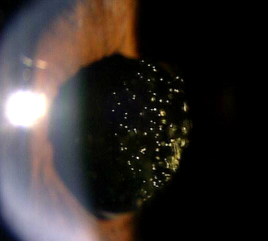
Crystalline deposits are also
known as “asteroid hyalosis”
ICD- 10 Diagnosis Codes:
H43.21–Crystalline deposits in vitreous body, right eye
H43.22–Crystalline deposits in vitreous body, left eye
H43.23–Crystalline deposits in vitreous body, bilateral
Title
Crystalline Deposits in Vitreous
Category
Disorders Of The Vitreous
Description
Crystalline deposits represent calcium-laden particles suspended within and attached to the structure of the vitreous body.
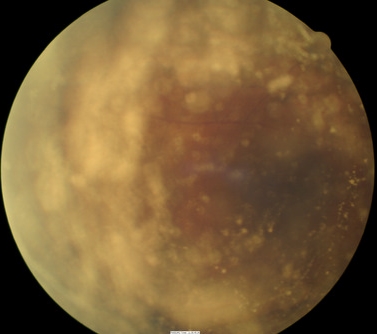
Asteroid hyalosis (Benson’s disease) is a benign condition and appears as multiple, discrete, yellow or yellow-white particles suspended in the vitreous, representing small calcium-laden lipids suspended within and attached to the hyaluronic acid framework of the vitreous body. They accumulate inferiorly and there are fewer bodies in the early stages.
Asteroid hyalosis is a primarily unilateral disorder that typically occcurs in patients over 60 and in men twice as often as women. Usually asymptomatic, in severe cases asteroid hyalosis can mildly affect visual acuity. Complaints of floaters are a rarity.
Even though the composition of the asteroid bodies is known, the exact genesis remains unclear. Current theories suggest that asteroid hyalosis results from aging collagen within the vitreous or a depolymerization of hyaluronic acid.
Sturctural Damage to the Eye
- No major structural damage
Functional Damage to the Eye
- No major functional damage
- Visual acuity may be mildly affected
The density of asteroid hyalosis does not correlate with visual dysfunction. If a patient presents with significantly diminished visual acuity, asteroid hyalosis is not to blame.
Patients with asteroid hyalosis and an unknown medical status require evaluation for the following systemic conditions:
- Diabetes
- Hypertension
- Hyperlipidemia
- Atherosclerotic vascular disease
 |
Fundus Photography
|
|
 |
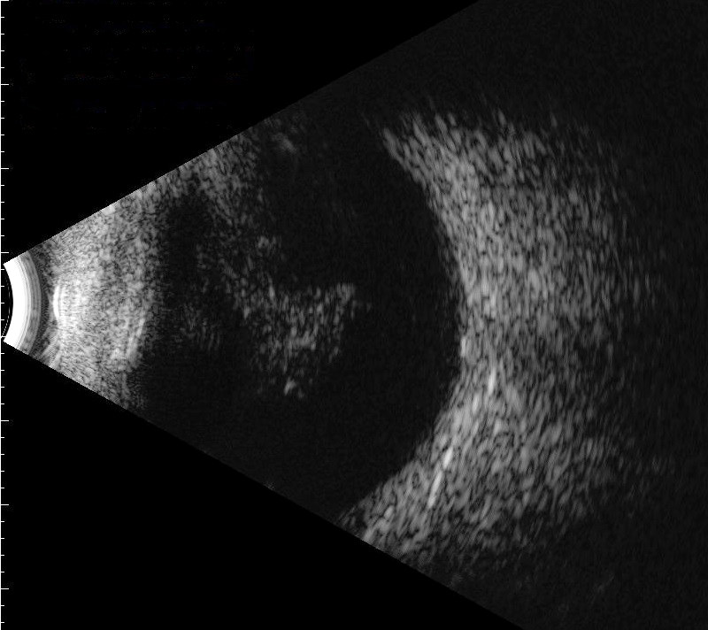
B-Scan Ultrasound
|
There is no classification system in place for asteroid hyalosis.
Synchisis Scintillans
Synchisis scintillans (cholesterol bulbi) is an extremely rare condition that occurs in severely diseased eyes. This condition also presents with refractile crystals in the vitreous, although these particles are composed of cholesterol. They are not attached to the vitreal framework, so they tend to settle out inferiorly after eye movement. Because this condition occurs in end-stage eye disease, pathologists rather than clinical optometrists or ophthalmologists typically make the diagnosis of synchisis scintillans.
Amyloidosis
Amyloidosis of the vitreous is also quite rare, and occurs typically after age 40. Patients characteristically demonstrate bilateral involvement with granular, strand-like opacities within the central vitreous. These membranes are anchored to the posterior lens surface in about half of patients. Small, yellow-white bodies dot the vitreal strands.
Usually, treatment is not necessary as asteroid hyalosis never leads to severe vision loss, and the mild symptoms occur rarely. Consider treatment only in patients who are also being managed for retinal disease (proliferative diabetic retinopathy, retinal tear or detachment).
Vitrectomy
Vitrectomy is surgery to remove some or all of the vitreous humor from the eye. Anterior vitrectomy entails removing small portions of the vitreous from the front structures of the eye — often because these are tangled in the intraocular lens or other structures. Pars plana vitrectomy is a general term for a group of operations accomplished in the deeper part of the eye, all of which involve removing some or all of the vitreous
1. Sowka JW, Gurwood AS, Kabat AG. Asteroid Hyalosis. Handbook of Ocular Disease Management. http://www.reviewofoptometry.com/cmsdocuments/2009/9/ro0409_handbook.pdf. Last accessed June 13, 2014.
2. Moss SE, Klein R, Klein BE. Asteroid Hyalosis in a Population: The Beaver Dam Eye Study. 2001 July. http://www.ncbi.nlm.nih.gov/pubmed/11438056. Last accessed June 13, 2014.
379.22
Crystalline deposits in vitreous
92250
Fundus photography
92225
Extended ophthalmoscopy
76512
B-Scan ophthalmic ultrasound
92133
Retinal laser scan (RNFL)
Occurrence
- Typically in patients over 60
Distribution
- Men twice as often as women
- In white patients, occurs in 1% – 2% of the population


 Print | Share