

 Print | Share
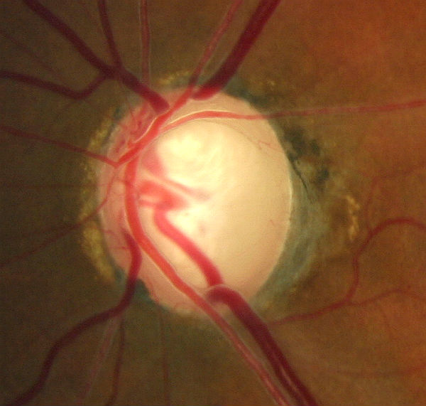
Advanced glaucomatous damage to optic nerve.
Case Report ID: 10
Title
Elevated-Tension Glaucoma; Advanced Damage
Category
Glaucoma (8)
Description
The case presents the diagnosis and treatment of elevated-tension open-angle glaucoma.
Glaucoma is an optic neuropaty showing distinctive changes in optic nerve morphology without associated pallor. The term “glaucoma” refers to a group of chronic, progressive optic neuropathies that have in common characteristic morphologic changes at the optic nerve and retinal nerve fiber layer.
The glaucomas are associated with the following clinical features:
- Aqueous outflow resrictions
- Unphysiologic intraocular pressure
- Abnormal ocular perfusion
- Abnormal rate of apoptosis
- Progressive retinal ganglion cell loss
- Characteristic changes in optic nerve anatomy
Case Report
- A 49-year-old black woman presented with a chief complaint of decreased vision
- Case history described a subjective decrease in vision over the past year
- The patient had been examined eight months earlier by another eye doctor and had received a prescription for bifocal eyeglasses
Conclusion
Although it may be very easy to conclude that this patient has glaucoma, the decisions regarding the problems associated with glaucoma and especially advanced glaucoma are very important and often difficult to make.
History of Present Illness
- Associated Signs and Symptoms: epiphora and ocular discomfort when reading
- Location: vision is worse in the left eye
- Duration: past one year
- Quality: n/a
- Context: “when I wake up – it takes a few minutes to focus”
- Severity: moderate loss of vision
- Timing: vision seems to be getting worse over time
- Modifiers: prescription eyeglasses eight months earlier – no improvement in vision
Review of Systems
- The patent reported that she was in excellent health and taking no medications
Past, Family and Social History
- Non-contributory
Best Corrected Distance Visual Acuity
- 20/20 in the right eye
- 20/50 in the left eye
Normal Examination Findings
- Mental status
- General medical observation
- Basic sensorimotor examination
- External examination
- Adnexal examination
- External ocular examination with biomicroscopy
Intraocular Pressure Measurements
- 43 mm Hg in the right eye
- 47 mm Hg in the left eye
Pupillary Examination
- Afferent pupillary defect in the left eye
Gross Visual Fields
- Confrontation testing revealed a severe constriction in the all meridians of gaze in the left eye
Ophthalmoscopy
- Cup-to-disc ratio of .60/.70 in the right eye
- Cup-to-disc ratio of .80/.90 in the left eye
- Vertical elongation of both optic cups
- Focal notch inferiorly and superiorly on the left optic disc
- Nasalization of the vascular tree in the left eye
- Rim/disc ratio < 0.1
 |
 |
.
Clinical Diagnosis
The clinical diagnosis is a determination based on the knowledge obtained from the patient’s medical history and from the results of the eye examination alone, without the benefit of diagnostic tests or procedures.
The patient’s clinical diagnosis is elevated-tension open-angle glaucoma based on the following clinical findings:
- Optic neuropathy in both eyes
- Elevated intraocular pressure measurements in both eyes
- Gross visual field defect in the left eye
- Relative afferent pupillary defect in the left eye
- Decreased visual acuity in the left eye
Treatment Plan
To gather the clinical information required to treat elevated-tension open-angle glaucoma, a diagnostic and treatment program is necessary and would include:
- Determination of different types of diagnoses
- Selection of one or more treatment options
Glaucoma is a disease that must be classified by type to be treated properly. After a clinical diagnosis has been determined, the diagnostic process continues with a process that involves the identification and exclusion of differential diagnoses. The differential diagnosis process allows the eye doctor to distinguish between two or more diseases with similar signs and symptoms by systematically comparing their signs and symptoms.
Differential Diagnoses
The first differential diagnosis that needs to be determined in this patient is glaucoma vs. some other retinal or neurological disease. The list of possible diseases would include any pathology that produces an abnormal optic nerve appearance, elevated intraocular pressures and associated visual field defects.
The following diseases share some of the clinical signs and symptoms of glaucoma; however, none of them has elevated intraocular pressure as a component of its clinical presentation.
-
Retinal detachment
-
Pituitary tumors and other neurological conditions
-
Ischemic optic neuropathy
-
Vascular disease
-
Drusen of the optic disc
-
Congenital disc anomalies
These conditions would not routinely be associated with significant elevation in intraocular pressure which strongly points to glaucoma as the correct etiology.
Ordering Diagnostic Tests
When additional clinical information is needed to complete the differential diagnostic process, diagnostic tests and procedures are performed. The performance of these tests and procedures leads to the completion of the differential diagnostic process while simultaneously initiating the treatment program.
Based on the clinical diagnosis of glaucoma, the following diagnostic tests and procedures were ordered at the conclusion of the eye examination.
- Refraction
- Retinal laser scan
- Visual field examination
- Gonioscopy
The decision to order and perform additional testing is totally based on the concept of medical necessity which can only be determined by the examining optometrist or ophthalmologist.
Refraction
- Measuring visual acuity is a method of evaluating functional vision loss
- Advanced glaucoma can produce central visual field defects which result in a loss of visual acuity
- There was no improvement in the left eye’s visual acuity after a subjective refraction was performed
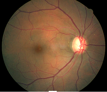
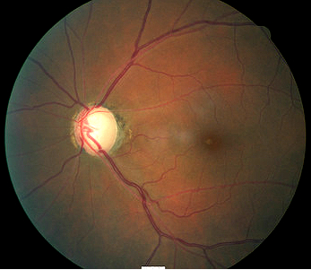
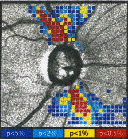
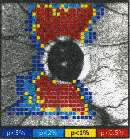
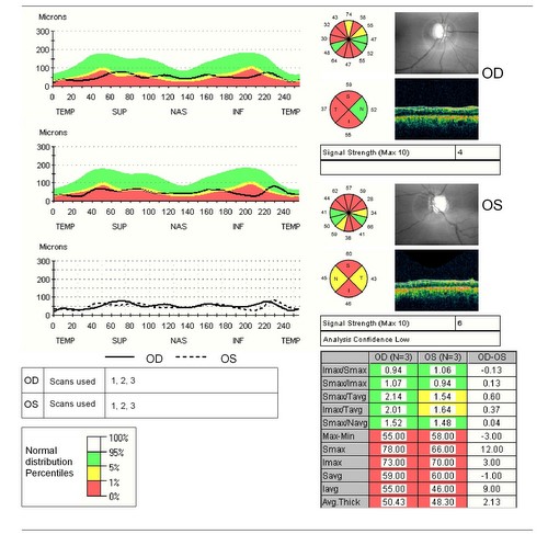
Retinal Laser Scan – Nerve Fiber Layer
- Measuring the average retinal nerve fiber layer thickness provides an overall assessment of retinal health
- The procedure can be accomplished by using the GDxVCC scanning laser polarimeter manufactured by Carl Zeiss Meditec as well as other scanning lasers
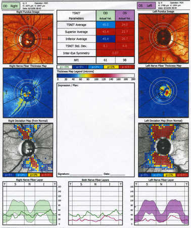 |
Scanning Laser Polarimetry
|
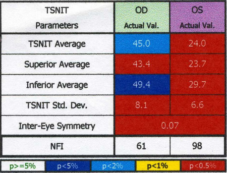 |
Inter-Eye Symmetryand the NFI Number
|
 |
 |
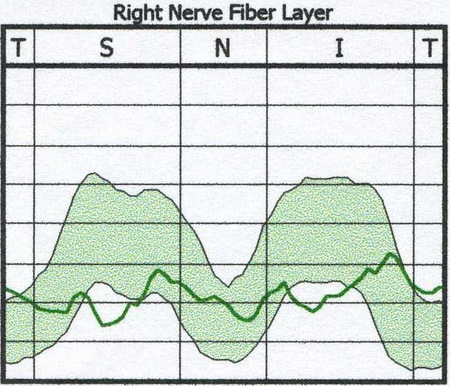
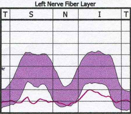
TSNIT Curve Profile Analysis
- The TSNIT Curve Profile Analysis is a linear representation of the scan showing the measured retinal nerve fiber layer thickness
- The slope and modulation of the patient’s profile should follow the normal “double-hump” distribution profile of the retinal nerve fiber layer
- In glaucoma, the superior and/or inferior retinal nerve fiber layer bundles are depressed or flattened
- Clinically significant asymmetry between the eyes is often found in patients with glaucoma
Right Eye
- Profile in the right eye shows flattening of the superior and inferior retinal nerve fiber layers
Left Eye
- Profile in the left eye is essentially flat – a finding that is characteristic of diffuse loss of the retinal nerve fiber layer
 |
 |
Retinal Laser Scan — Nerve Fiber Layer
 |
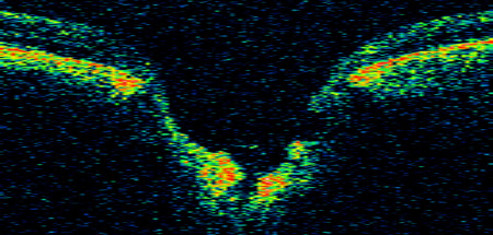
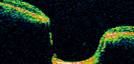
Optical Coherence Tomography
|
Retinal Laser Scan — Optic Nerve Head
- xxxxx
- xxxxx
 |
 |
Visual Field Examination
- Automated threshold perimeters meaure the visual field by ploting the threshold luminance value of the patient in various locations in the visual field
- The luminance of the light stimulus is represented by non-specific units of measurement called deciels (dB)
- Glaucoma produces several changes in the visual field, and one of these changes occurs as a widespead, non-descript loss of retinal sensitivity
- In Tranquair’s “Hill of Vision” concept, loss of retinal sensitivity represents a reduction in the height of the hill
- The diffuse loss of retinal sensitvity should be considered highly diagnostic of glaucoma when it is asymmetric and correlates with asymmetric changes in intraocular pressure or disc appearance
- In most cases, the loss of sensitivity occurs in characteristic patterns and locations (e.g., nasal step, arcuate scotoma, paracentral scotoma) that often correlate with changes in the optic nerve and/or retinal nerve fiber layer.
 |
 |
Automated threshold perimeters characterize specific parameters of the overall visual field status by the use of numbers called Global Indices. One of the indices, Mean Deviation (MD), expresses the raw data generated by the instrument.
A visual field defect can be classified as mild, moderate or advanced based upon an abnormal Mean Deviation.
- Mild visual field defect = 0 through -5.99 dB
- Moderate visual field defect = -6.00 dB through -11.99 dB
- Advanced visual field defect = anything above -12.00 dB
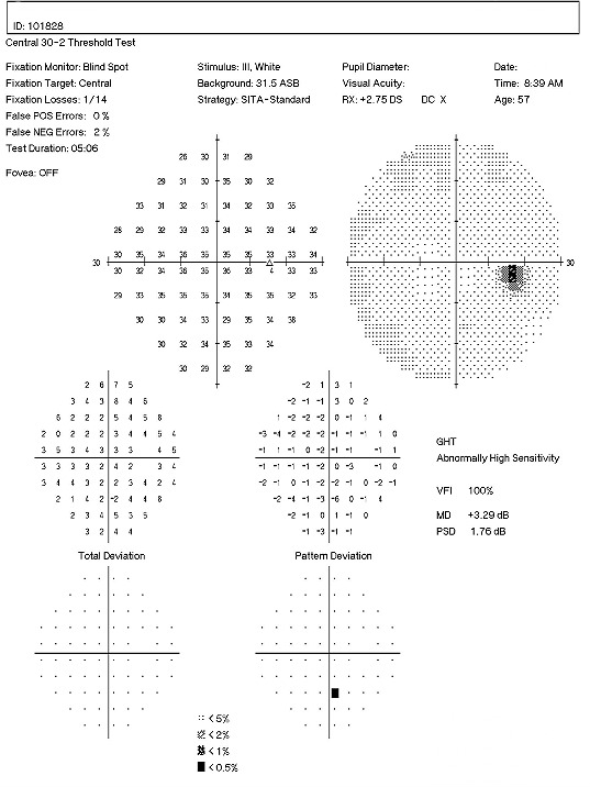
As represented by the Mean Deviation of +3.29 dB, the visual field of the right eye demonstrated a mild loss of retinal sensitivity.
- Scattered paracentral scotomas were found throughout the central visual field in the right eye
- Paracentral scotomas are visual field defects characterized by small, isolated areas of reduced retinal sensitivity
- They are typically centered just beyond the central 10 degrees of fixation but usually within 20 degrees of fixation
- Paracentral scotomas occur in about 70% of patients with early glaucomatous damage to the visual system
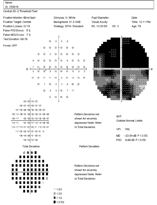
As represented by the Mean Deviation of -23.54 dB, the visual field of the left eye demostrated a severe loss of retinal sensitivity.
- The examination revealed a constriction of the isopters to the fixation point inferiorly
- A dense arcuate scotoma was found in the inferior visual field of the left eye
- A moderate arcuate scotoma was found superiorly
- Arcuate scotomas are visual field defects that occur when paracentral scotomas expand or coalesce with other paracentral defects
- The presence of this type of defect represents moderate-to-advanaced glaucomatous damage to the visual system
The results of the examination revealed definite glaucomatous visual field defects in each eye. The loss of retinal sensitivity should be considered highly diagnostic of glaucoma when it is asymmetric and correlates with asymmetric changes in intraocular pressue, retinal nerve fiber layer appearance or optic disc appearance. Because the visual field defects in each eye agree with measurable, observable structural damage to the eye, the clinical diagnosis of elevated-tension open-angle glaucoma is confirmed.
Gonioscopy
- The anatomical configuration of the iris and angle is important in the pathogenesis of certain forms of glaucoma
 |
Gonioscopy Examination Results
There is no apparent obstruction of the anterior chamber angle or outflow mechanism. |
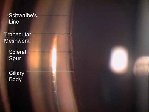 |
Angle Grading System
This patient’s angles were determined to be Grade 4 in all quadrants. |
All of the diagnostic test results confirmed the initial diagnosis of elevated-tension open-angle glaucoma. Additional diagnostic tests for glaucoma such as serial tonometry, provocative glaucoma testing, or extended ophthalmoscopy were not medically necessary on this patient’s initial visit. Photodocumentation of the optic nerve should be performed at some follow-up visit.
According to Current Procedural Terminology, when eye doctors perform ophthalmological examinations, the complexity of medical decision-making is not separated from the examining techniques used. As a guideline to assist eye doctors in enhancing their medical decision-making skills, consider that the complexity of medical decision-making involves three components.
The first component concerns the number of possible diagnoses and treatment options that must be considered. Modern glaucoma diagnosis involves the determination of structural damage to the eye and/or functional loss to the visual system. Once these assessments are made, the glaucoma must be classified by type.
The second component concerns the amount and complexity of medical records and diagnostic tests that have to be obtained, reviewed and analyzed. In addition to an eye examination, this visit required the review and analysis of a subjective refraction, a retinal laser scan, a threshold visual field examination, and a gonioscopic examination of the anterior chamber.
Third, the complexity of medical decision-making is affected by the risk of significant complications and/or morbidity associated with advanced glaucoma and the risks involved in any treatment options. This patient’s glaucoma was classified as a severe problem where the risk of total vision loss without treatment was significant. In addition, because the treatment plan would involve chronic pharmaceutical treatment with the potential for ocular and systemic side effects, the pharmacology complications had to be considered when deciding on the initial treatment options.
Treatment Guidelines
To begin the treatment of glaucoma, intraocular pressure must be lowered and an initial target pressure range must be established. A general guideline is to base the initial pressure range on the category of damage to the visual system.
- Mild damage needs a 20-30% reduction in pre-treatment intraocular pressure as the initial target pressure range
- Moderate damage needs a 30-40% reduction in pre-treatment intraocular pressure as the initial target pressure range
- Advanced damage needs a 40-50% reduction in pre-treatment intraocular pressure as the initial target pressure range
Due to the advanced glaucomatous damage in the left eye, a 40-50% reduction in the pre-treatment intraocular pressure was selected as the initial target pressure range.
Doctors should always remember that the goal in glaucoma therapy is preventing future or additional vision loss, not chasing some arbitrary pressure range.
Treatment Program
Modern glaucoma therapy generally utilizes prostaglandins as the primary therapy. These medications typically have the greatest efficacy. The first goal of treating glaucoma is to achieve the lowest possible intraocular pressure on a single agent. Monotherapy with prostaglandins improves patient compliance, may decreases costs, and improves the safety profile and tolerability of using topical medications. These drugs reduce intraocular pressure by increasing uveoscleral outflow. Intraocular pressure reduction averages almost 30%, sometimes more and sometimes minimal to no effect.
Clinically significant conjunctival hyperemia is the most common side effect of prostaglandin therapy. Another possible ocular side effect of prostaglandins includes changes in iris color after several months of use. In these patients, the drug produces an increase in the amount of iris stromal pigmentation. Over time, eyelashes can lengthen, thicken, and darken – a side effect known as hypertrichosis.
The only serious ocular side effect is a 3% increased risk of developing cystoid macular edema. A history of uveitis is a relative contraindication, as prostaglandins produce an increased risk of reactivating the inflammation. The systemic side effects of topical prostaglandins are minimal.
Avoiding medicolegal liability if complications arise is an important part of a modern ophthalmic practice. This avoidance is best accomplished by making sure that you have obtained the patient’s informed consent to treat their glaucoma. Once you have made the patient fully aware that using topical glaucoma medicine involves the risks of side effects, you can proceed with the treatment plan.
After obtaining the patient’s informed consent to treat her glaucoma, Travatan Z ophthalmic solution was prescribed 1 drop at bedtime in both eyes. A target pressure was set at OD 21 mm Hg and OS as low as possible. It would be typically unrealistic to expect any monotherapy to achieve the desired target endpoints, although this can rarely occur with prostaglandin therapy.
Next examination scheduled for 2 weeks. Full pressure reduction with prostaglandins is typically not achieved until 30 days but Travatan Z has been shown to reach near full potential in as little as two weeks.
Discussion
This case demonstrates several key aspects of glaucoma diagnosis and treatment. First, despite the advanced damage to the left eye, there was no ocular emergency and no need for a referral to a glaucoma sub-specialist at this time. This patient had been functioning with glaucoma for likely many years. Another few days or weeks without treatment would probably not change the course of the disease.
Second, this patient’s clinical history supports the modern understanding of the pathophysiology of glaucoma. We know that glaucoma is an optic neuropathy. Structural damage to the retinal nerve fiber layer is usually the first milestone in the natural history of glaucoma. The loss of retinal nerve fibers is closely followed by changes in the appearance of the optic disc. Functional loss, which is documented by abnormal visual field testing, abnormal visual evoked potential testing or loss of visual acuity, usually occurs later in the natural history of the disease. In this case, there is advanced functional damage to the left eye.
On initial examination, the damage to the optic disc was advanced in the left eye, and its prognosis for continued good vision ten or twenty years from now is poor. Without significant therapeutic effect from medication, it is likely this left eye will require laser trabeculoplasty (SLT). The advanced damage and fragile nature of the remaining nerve fibers in the left optic nerve is likely to eliminate consideration of the risks associated with a filtering procedure. The right eye, while having less damage to the retinal nerve fibers and only mild visual field defects, had a very abnormal retinal laser scan and elevated IOPs. The abnormal retinal laser scan suggests that this patient’s right eye is in the interval between the measurement of structural damage from glaucoma and the measurement of functional vision loss from glaucoma. This gap, up to five years in some patients, often delays and confuses eye doctors in trying to diagnose glaucoma or to assess its severity.
Advanced Glaucoma Intervention Study
Although the Advanced Glaucoma Intervention Study found that intraocular pressures of 12 mm Hg or below were best for patients with advanced glaucoma, this target range is typically not necessary during the initial stage of treatment and not essential in this patient’s case. On the patient’s second visit two weeks later, intraocular pressure measured 24 mm Hg in each eye. This patient’s initial response to treatment demonstrates the efficacy of prostaglandins: a greater than 40% reduction in pre-treatment intraocular pressure achieved on monotherapy in two weeks. It is unlikely this pressure will be sufficient to protect the left eye so careful monitoring for any continued progression is warranted.
Baltimore Eye Study
This patient is a reminder that the findings from the Baltimore Eye Study are still true. Although this patient was initially examined in 2006, some of the problems in diagnosing glaucoma that were revealed by the study years ago are still present. One of the findings from the Baltimore Eye Study is that glaucoma can be hard to diagnose. The study revealed that 50% of all people found to have glaucoma during the study had seen an eye doctor within the past year and were unaware that they had glaucoma. In this case, the patient had been examined by another eye doctor eight months earlier and was not diagnosed with glaucoma.
Based on the patient history, the nature of the presenting problem, and my own clinical judgement this patient needed an evaluation of the complete visual system.
- Perform the eye examination that is medically necessary
- Provide the diagnostic tests or services that are medically necessary
- Properly document the services provided
- Code from the documentation
- Report the services to the payor
| Diagnosis Code | Procedure Code | Modifier | Quantity | Payor | Amount Allowed |
| H40.1133 - Primary open-angle glaucoma, bilateral, severe stage | 92004 - Medical eye examination | 1 | Self | 150.00 | |
| H40.1133 - Primary open-angle glaucoma, bilateral, severe stage | 92015 - Refraction | 1 | Self | 25.00 | |
| H40.1133 - Primary open-angle glaucoma, bilateral, severe stage | 92133 - Retinal laser scan | 1 | Self | 50.00 | |
| H40.1133 - Primary open-angle glaucoma, bilateral, severe stage | 92083 - Visual field examination | 1 | Self | 75.00 | |
| H40.1133 - Primary open-angle glaucoma, bilateral, severe stage | 92020- Gonioscopy | 1 | Self | 40.00 | |
| Total | $340.00 |
H40.1133
Primary open-angle glaucoma,
bilateral, severe stage
365.11
Primary open-angle glaucoma
92015
Refraction
92020
Gonioscopy
92083
Visual field examination
92133
Retinal laser scan
92250
Fundus photography
95930
Visual evoked potential
92275
Electroretinography
92225
Extended ophthalmoscopy
76514
Corneal pachymetry
76513
Anterior segment ultrasound
92132
Anterior segment imaging
92100
Serial tonometry