

 Print | Share
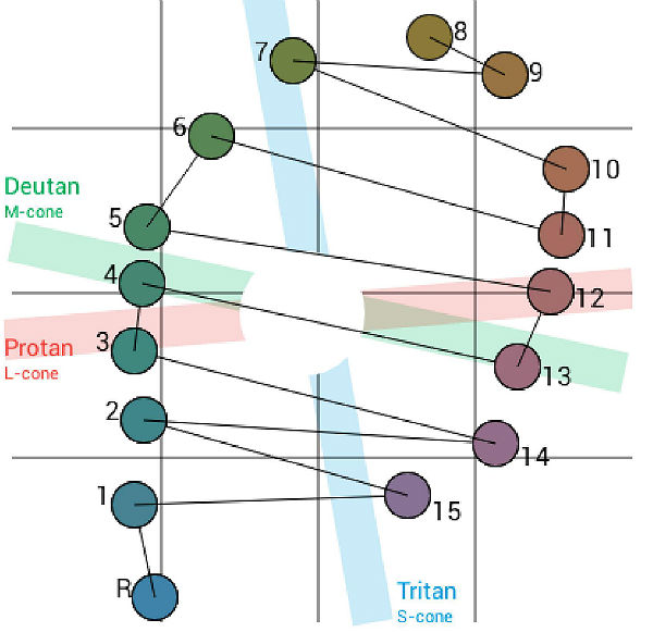
The color confusion lines of genetic defects have an orderly pattern and are almost parallel to each other
ICD-10 Diagnosis Code:
H53.53–Deuteranomaly
Title
Deutan Color Vision Defect
Category
Visual Disturbances
Description
A genetic red-green color vision deficiency resulting from inherited green cone pigment abnormalities.
Color is perceived because objects selectively absorb certain wavelengths of light while transmitting the other wavelength; and, the object will take on the color of the wavelength of light it transmits.
The perception of color is defined by three variables:
- Hue — the dominant wavelength of the light the object transmits
- Saturation — the absence of white
- Brightness — the intensity of the color
The retina contains two types of photoreceptors:
- Rods — used for vision in low light
- Cones — used for vision in bright light and for color vision
Color vision is a function of the retinal cones and each photoreceptor cell contains a specific type of photopigment. Normal color vision is trichromatic and is based on three types of cones that are maximally sensitive to light at certain wavelengths.
- Blue cones — sensitive to light rays of short wavelength (approx. 420 nm)
- Green cones — sensitive to light rays of middle wavelength (approx. 530 nm)
- Red cones — sensitive to light rays of long wavelength (approx. 560 nm)
People with an inherited deutan color vision deficiency produce decreased sensitivity to perception of the green cone pigment.
Genetic deutan color vision defects appear in early infancy and once fully expressed, the defect remains stable throughout life. The defects are divided into subclasses based on severity and the type of missing or anomalous color cone receptor.
Structural Damage to the Eye
- None, red-green color vision defects are not accompanied by any other ocular abnormality
- Retina appears normal
- Deutanomaly, normal blue and red cones plus anomalous green-like cones (5% of white males)
- Deutanopia, blue and red cones only; no functional green cones (1% of white males – more severe form of the disease)
Functional Damage to the Eye
- Decreased color vision
- Most people with deutanomalous defects have no problem naming colors
People with deutan defects may be excluded from a variety of industrial, marine, air, rail, and military occupations that require the ability to distinguish red and green colors. Although people with mild defects may be able to discriminate color, they may fail the strict requirements of color discrimination on extended color vision examinations.
The goal of the diagnostic evaluation in a patient with an deutan color vision deficiency is to accomplish the following:
- Differentiate whether the color vision defect is genetic or acquired
- Determine if the color vision defect is unilateral, asymmetric, or transient
- Identify the primary condition that is producing the color vision defect
- Prescribe a treatment program
Patient History
Deutanopia
- Most people with deutanopia perceive the visible spectrum as lacking red, orange, green, blue, and cyan
- There is market difficulty in selecting colored articles, materials, and foods
Deutanomaly
- Most people with deutanomaly can perceive colors but color saturation is weakened
- Color discrimination deficits vary widely in severity
Normal Color Vision vs. Abnormal Color Vision
- Patients with deutanomaly are not color blind, they have a color matching deficit

Clinical Appearance of the Retina
There are no retinal changes associated with a genetic deutan color vision deficiency.
DIAGNOSTIC TESTS
Color Vision Examination
Tests of Color Saturation Perception
The most common test to identify genetic deutan color vision defects is Pseudoisochromatic plates. Of these, Ishihara pseudoisochromatic plates is the most recognized and utilized. Ishihara plates are composed of embedded characters and people with normal color vision will see most or all of the characters. Because people with genetic deutan color vision defects cannot distinguish colors along the confusion lines, they will not be able to see some of the embedded characters in the plates.
Extended color vision testing divides people into two groups:
- The first group consist of people with normal color vision and slight color deficiency
- The second group consists of people with moderate or severe color deficiency
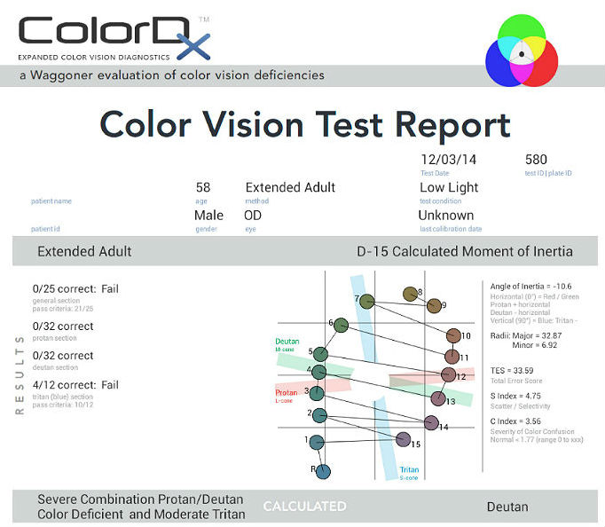
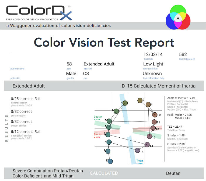
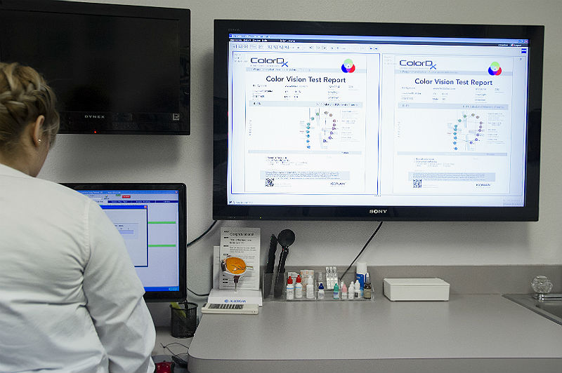
The most common test performed in clinical practice is the Farnsworth D15 and the procedure can be accomplished using Konan’s ColorDx software. The test consists of fifteen colored bars and one fixed bar. The hue of each bar has been chosen so that adjacent bars have approximately equal hue differences. When the bars are arranged in order they form a hue circle. As a result, errors in hue discrimination can be made across the hue circle.
 |
 |
|
 |
ColorDx — Color Vision Diagnostic
|
|
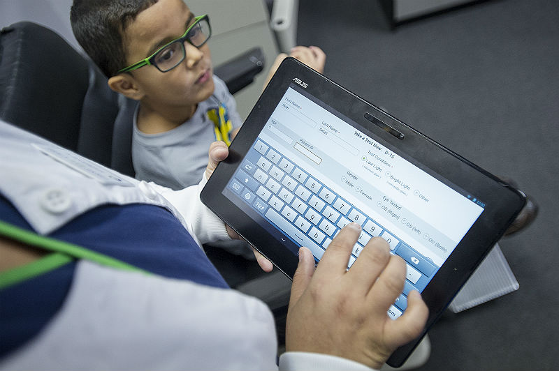
ColorDx — Pediatric Testing
|
 |
|
 |
Patients with normal color vision usually make no error but may make one or two minor transposition errors, as do those with mild color vision deficiency. Patients with more advanced color deficiency make some or more of the following errors:
|
Acquired Color Vision Deficiency
Several eye diseases and clinical conditions affect a person’s color vision. These acquired color vision deficiencies are characterized by a reduction in a person’s ability to discriminate between different wavelengths of visible light.
In contrast to genetic color vision defects, which are always bilateral, acquired color vision deficiencies can be monocular.
Common conditions that can affect a person’s color vision include the following:
- Normal age-related deterioration in chromatic discrimination ability
- Yellowing caused by cataract results in a loss of hue discrimination
- Diseases that result in a loss of foveal function
- Optic nerve disease
- Retinal dystrophies
- Neurologic disease
- Neurologic injury
- Visual field defects
- Common drugs and substances
The goals of treating a patient with a deutan color vision deficiency include the following:
- Differentiate between genetic and acquired color vision deficiency
- Differentiate between deutanomaly and deutanopia (e.g., anomalous trichromat vs. dichromat)
- Prescribe a program to treat the manifestations associated with deutan color vision defects
Tinted Lenses
Specialized tinted lenses that filter specific wavelengths of light may minimally improve color discrimination. The tint selection process requires that the patient select a preferred tint from a group of colors across the visible spectrum.
- ChromaGen — FDA marketing clearance for use in red-green color deficiencies
1. Karpecki P, Shechtman D. Color Me Curious. RevOptom. 15 Feb 2013. http://www.revoptom.com/content/c/39720/dnnprintmode/true/?skinsrc=%5Bl%5Dskins/ro2009/pageprint&containersrc=%5Bl%5Dcontainers/ro2009/simple. Last accessed May 31, 2014.
2. Deeb S, Motulsky A. Red-Green Color Vision Defects. 29 Sept 2011. http://www.ncbi.nlm.nih.gov/books/NBK1301/. Last accessed May 31, 2014.
3. Pacheco-Cutilla M, Edgar D. Acquired colour vision defects in glaucoma — their detection and clinical significance. Br J Ophthalmo. 1999. http://bjo.bmj.com/content/83/12/1396.full. Last accessed May 31, 2014.
4. Cole B. Assessment of inherited colour vision defects in clinical practice. 11 Apr 2007. http://onlinelibrary.wiley.com/doi/10.1111/j.1444-0938.2007.00135.x/full. Last accessed May 31, 2014.
5. Ivan DJ. Ophthalmology. Rayman’s Clinical Aviation Medicine 5th Ed. 2013; (235-292).
6. Pubmed Health. Color Blindness. 2011 June 1. http://www.ncbi.nlm.nih.gov/pubmedhealth/PMH0001997/. Last accessed July 26, 2014.
7. Evaluation of Acquired Color Vision Deficiency in Glaucoma Using the Rabin Cone Contrast Test. Invest Ophthalmol Vis Sci. 2014 Aug 28;55(10):6686-90. http://www.ncbi.nlm.nih.gov/pubmed/25168899. Last accessed May 18, 2015.
368.52
Deutan defect
92283
Color vision examination
Occurrence
- 1% of white males have deuteronopia
- 5% of white males have deuteranomaly
Distribution
Genetic red-green color vision defects are common.
- 8% of males — northern European descent
- 3% – 4% of males — African ancestry
- 3% of males — Asian ancestry
- 0.5% of females — northern European descent
Risk Factors
- Persons of northern European descent