

 Print | Share
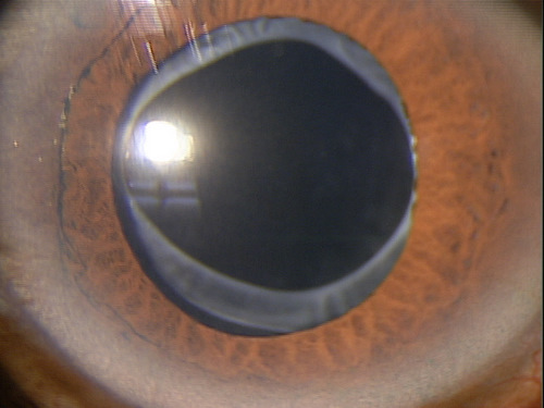
Pseudophakia
ICD-10 Diagnosis Code:
Z96.1 — Presence of intraocular lens
Title
Lens Replaced by Other Means
Category
Organ Or Tissue Replaced By Other Means (1)
Description
Pseudophakia is the term used to describe the replacement of a partial or complete opacity on or in the lens or capsule of one or both eyes with an artificial one.
| Pseudophakia is the term used to describe the replacement of a partial or complete opacity on or in the lens or capsule of one or both eyes with an artificial one. | 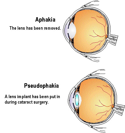 |
Structural Damage to the Eye
- Acquired loss of endothelial cells secondary to intraocular lens implant surgery
- Cystoid macular edema can result from intraocular lens implant surgery
Functional Damage to the Eye
- Secondary cataracts can form on the intraocular lens
- Blurred vision due to secondary cataracts on the intraocular lens
- Corneal edema secondary to loss of endothelial cells after intraocular lens implant surgery
The main goal of the diagnostic evaluation in a patient with pseudophakia is to accomplish the following:
- Determine if the intraocular lens is centered
- Assess if secondary opacities are present
- Determine if iatrogenic endotheliopathy has occurred
- Evaluate the macula with OCT technology to determine if postoperative vitreoretinal pathology is present
- Determine a treatment plan
Patient History
Patients will present with any of the following signs and symptoms:
- No visual complaints
- Blurred and/or distorted vision
- Difficulty seeing in bright light
External Ocular Examination with Biomicroscopy
 |
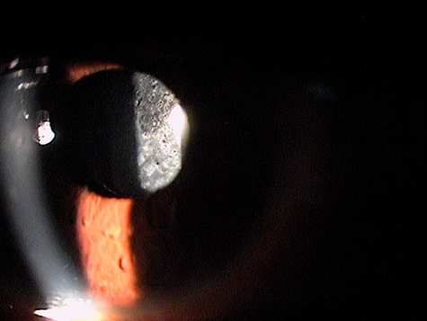
Clinical Appearance of the Posterior Chamber
|
|
 |
Clinical Appearance of the Posterior Chamber
|
|
 |
Clinical Appearance of the Posterior Chamber
|
|
 |
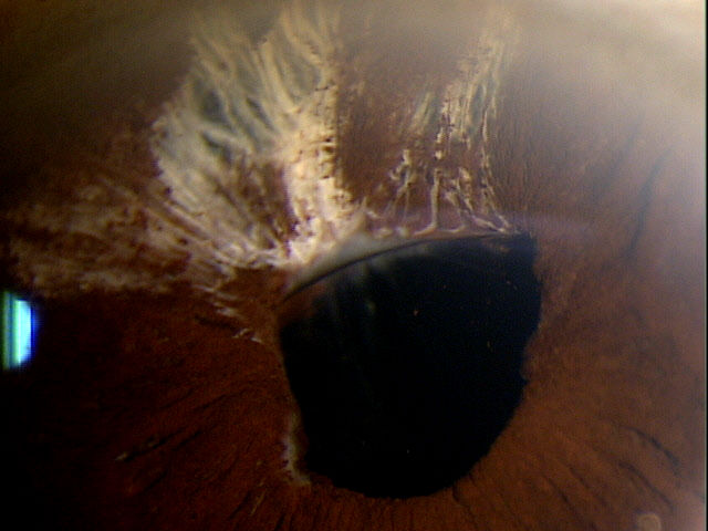
Clinical Appearance of the Anterior Chamber
|
DIAGNOSTIC TESTS
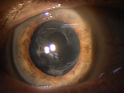
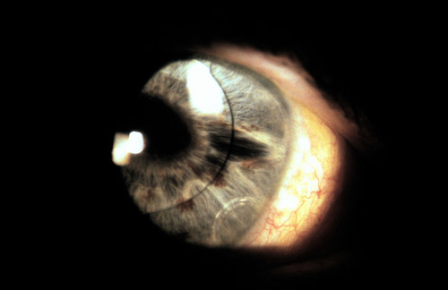
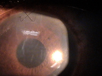
External Ocular Photography
- Document the severity of any secondary opacities
- Document the progress or lack of progress of the opacities
- Help determine a treatment plan
 |
Pseudophakia
|
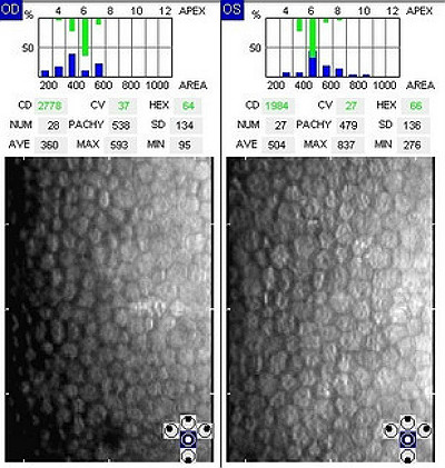
Specular Endothelial Microscopy
- Earlier and more accurate diagnosis of iatrogenic endotheliopathy
- To evaluate unexplained loss of visual acuity after surgery
- Document the progress or lack of progress of a corneal endotheliopathy
- To help plan a treatment program
- To document the delivery of medical treatment
- To document the response to treatment
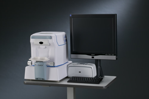 |
Konan Specular Microscope
|
|
Specular Microscopy — Three weeks Pre-OP
|
 |
|
Pseudophakia — Two weeks Post-OP in the right eye
|
 |
|
Specular Microscopy — Two weeks Post-OP
|
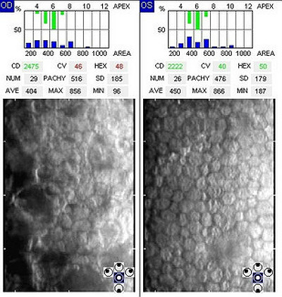 |
Retinal Laser Scan
- Earlier and more acurate diagnosis of retinal disease
- To evaluate unexplained loss of visual acuity after surgery
- Document the progress or lack of progress of vitreomacular traction syndrome
- Help plan a treatment program
- To document the delivery of medical treatment
- Document the response to treatment
- Measure the effectiveness of therapy
- Determine the need for ongoing therapy
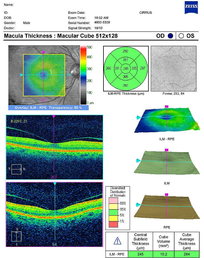 |
Optical Coherence Technology — Three weeks Post-OP
|
|
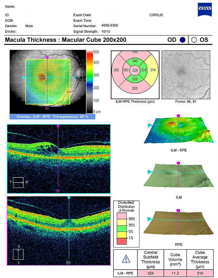 |
Optical Coherence Technology — Four months Post-OP
|
There is no classification system in place for pseudophakia.
None.
Optical Treatment
- Prescription eyeglasses
- Prescription contacts
Surgical Treatment
- YAG laser capsulotomy
- Refractice surgery for uncorrected postoperative refractive error
1. Graham R. Aphakic and Pseudophakic Glaucoma. 2 Sept 2014. http://emedicine.medscape.com/article/1207170-overview. Last accessed November 16, 2014.
2. Prevalence of Cataract and Pseudophakia/Aphakia Among Adults in the United States. Arch Ophthalmol. 2004;122(4):487-494. http://archopht.jamanetwork.com/article.aspx?articleid=416230. Last accessed November 16, 2014.
V43.1
Lens replaced by other means
92025
Corneal topography
92015
Refraction
92136
Ophthalmic biometry by A-scan with intraocular lens power calculation
92286
Specular endothelial microscopy
Occurrence
The prevalence of pseudophakia is linked to the number of senile cataracts.
- Senile cataracts are found in almost 66% of the population over 75 years old
- The prevalence is also linked to the number of congenial disorders that require cataract extraction
Distribution
- No preference for males or females
Risk Factors
- Age
- Trauma
- Congenital factors causing lens to be remove at a young age